Disease code
The International Classification of Diseases has assigned each type of disease its own code. According to ICD-10, umbilical hernia has an international code of K42. This is the main code, but there are also subspecies that are also assigned a special code.
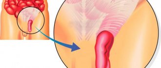
An umbilical hernia is a protrusion in the area where the navel is located. Anatomically, the hernia has:
- hernial orifice;
- hernial sac;
- hernial contents (usually intestinal loops).
What symptoms indicate that a patient has this disease? A hernia located in the navel area may not show any symptoms of its presence for quite a long time. The first symptoms of the disease can be observed when the formation is infringed.
When there are no symptoms yet, the following signs may indicate the presence of a formation:
- visible protrusion in the navel area;
- the protrusion has a soft-elastic consistency;
- when the disease is in the very initial stage, the protrusion can be easily reduced (such hernias are called easily reducible);
- The size of the hernial protrusion can vary depending on how long the hernia has been present.
If education is impaired, the course of the disease changes quite dramatically. Symptoms such as:
- severe pain in the area of protrusion of the umbilical hernia;
- there is a violation of stool;
- in some cases, an increase in body temperature may be observed;
- attacks of nausea or vomiting may occur;
- belching may have a rotten smell;
- weakness appears and appetite is lost.
This condition is dangerous for the patient, because there is a possibility of developing acute intestinal obstruction.
Causes of the disease
For an umbilical hernia to form, several factors must combine. This is an innate factor and acquired during life. The congenital factor is associated with a certain anatomical structure of the navel area. And acquired factors include an increase in intra-abdominal pressure, which causes the abdominal organs to protrude.
Factors that are considered congenital are:
- weak umbilical ring;
- congenital anatomical defect;
- connective tissue dysplasia;
- weak muscles of the anterior abdominal wall.
Factors that arise during a person’s life may be:
- cough (this especially applies to cough that occurs with chronic bronchitis or in those who smoke);
- constipation;
- some types of professions (for example, musicians who play wind instruments);
- persons engaged in heavy physical labor.
Diagnosing the presence of an umbilical hernia is not so difficult. To do this, you should conduct a particularly thorough examination, for which you will need to visit a specialist.
All other diagnostic measures are aimed at excluding possible complications of such a disease. To understand whether a patient has this formation, he needs to undergo a series of diagnostic procedures, such as:
- Ultrasound of the abdominal organs.
- X-ray examination. It is also necessary to add x-rays using contrast agents.
- General urine analysis. Such a study can show the inflammatory process that occurs against the background of strangulation of the umbilical hernia.
If treatment is untimely, there is a possibility that various complications may arise, such as:
- the appearance of intestinal obstruction;
- peritonitis;
- infectious-toxic or painful shock;
- coma state.
All this suggests that treatment should not be delayed under any circumstances. At the slightest suspicion of an umbilical hernia, the patient should immediately consult a doctor and undergo the necessary examinations.
How is an umbilical hernia treated?
Umbilical hernia is treated mainly surgically. Expectant management is used only in newborns and children who have not yet turned one year old.
This tactic is as follows:
- it is necessary to observe with particular frequency and dynamics the condition of the umbilical ring;
- You should wear special devices that will perform a support function.
Hernias are surgically removed only in adults or those children in whom expectant management has not led to any result.
The operation consists of several steps. First, the hernial sac is isolated. The contents of the hernial sac are returned to the abdominal space. Eliminate hernial orifices.
Using a special mesh, the hernial orifice is strengthened. Typically, during such an operation, the patient is given local anesthesia and is prohibited from lifting heavy objects for six months after the operation. Then you are allowed to return to your previous lifestyle.
To prevent the formation of an umbilical hernia, it is important to know what preventive measures should be followed. If a person is at risk and has an increased likelihood of developing an umbilical hernia, he should be examined by a surgeon. This must be done at regular intervals so that any pathology can be detected as quickly as possible.
Examinations should include x-ray and ultrasound examinations. In addition, the following recommendations should be followed:
- treat cough in a timely manner;
- fight constipation;
- for obesity, it is recommended to normalize body weight;
- stop smoking;
- You should not lift or carry heavy objects.
If the patient is nevertheless diagnosed with an umbilical hernia, then he is recommended to adhere to a certain lifestyle.
It is recommended to limit heavy lifting. You should wear a special suspensor, which prevents the protrusion of the hernial contents and its subsequent strangulation.
Treatment methods
In children from birth to 1 year, active expectant treatment is used, which includes:
- constant monitoring of the condition of the navel;
- use of support bandages;
- operation if containment measures are ineffective.
In adults and older children, the only method of effective treatment for umbilical hernia is surgery. With its help, the surgeon isolates the hernial sac and returns its contents back into the abdominal cavity. Then a special mesh is installed to restrain the hernial orifice. The operation is performed under local or general anesthesia, depending on the complexity of the patient’s condition.
The rehabilitation period is 6 months, the first weeks must be kept in bed.
If a hernia occurs in parallel with multiple malformations in a newborn child, the prognosis will be unfavorable. Early surgery or restraining therapy helps eliminate the hernia without recurrence. Adults tolerate surgery well; in the absence of associated complications, the risk of relapse is unlikely.
Strangulated umbilical hernia code according to ICD 10
ICD-10 code K42
You are here: Home > ICD-10 > K00-K93 > K40-K46
ICD-10 code K42 for Umbilical hernia
Includes: periumbilical hernia
Excludes: omphalocele (Q79.2)
ICD-10
ICD-10-CM 10th Revision 2016
ICD-10-GM ICD-10 in Germany
ICD-10 ICD-10 in Russian
ICD-10 International Statistical Classification of Diseases and Related Health Problems 10th Revision
ICD-10
ICD-10 is the 10th revision of the International Statistical Classification of Diseases and Related Health Problems (ICD), a medical classification list by the World Health Organization (WHO).
It contains codes for diseases, signs and symptoms, abnormal findings, complaints, social circumstances, and external causes of injury or diseases.
The Anatomical Therapeutic Chemical (ATC) Classification System is used for the classification of active ingredients of drugs according to the organ or system on which they act and their therapeutic, pharmacological and chemical properties.
It is controlled by the World Health Organization Collaborating Center for Drug Statistics Methodology (WHOCC).
The defined daily dose (DDD) is a statistical measure of drug consumption, defined by the World Health Organization (WHO).
It is used to standardize the comparison of drug usage between different drugs or between different health care environments.
An umbilical hernia is a surgical pathology caused by the exit of the abdominal organs beyond the anterior wall through a characteristic opening.
In all medical institutions, umbilical hernia according to ICD 10 has code K42 , which determines diagnostic methods, treatment tactics and patient management, as well as preventive measures. The code assumes several types of surgical disease, which determines the second digital value after the point, namely the following:
- the exit of the peritoneal organs through the umbilical ring, which causes the presence of obstruction, but is not complicated by a gangrenous course, has code K42.0;
- a hernial formation in the navel with the development of signs of gangrene is designated by code K42.1;
- protrusion of a hernial sac of unspecified etiology through the umbilical opening without signs of obstruction and necrosis defines code K42.9.
The classification characterizes each individual case.
Complications
The main complication of a hernia of various locations and sizes is its strangulation.
However, a strangulated umbilical hernia, along with a femoral or identical defect of the white line of the abdomen, is much less common than an inguinal hernia, which has a maximum prevalence among the adult population. In most cases, unilateral infringement is noted , which somewhat reduces the rate of increase in the symptoms of gangrene and general intoxication.
Save the link, or share useful information on social media. networks
Diagnosis with code K42 includes 3 clarifying diagnoses (ICD-10 subheadings):
Chain in classification:
The diagnosis also includes: peri-umbilical hernia
The diagnosis does not include: – omphalocele (Q79.2)
mkb10.su - International classification of diseases, 10th revision. Online version 2021 with disease search by code and decoding.
Strangulated umbilical hernia code according to ICD 10
ICD-10 code K42
You are here: Home > ICD-10 > K00-K93 > K40-K46
ICD-10 code K42 for Umbilical hernia
Includes: periumbilical hernia
Excludes: omphalocele (Q79.2)
ICD-10
ICD-10-CM 10th Revision 2016
ICD-10-GM ICD-10 in Germany
ICD-10 ICD-10 in Russian
ICD-10 International Statistical Classification of Diseases and Related Health Problems 10th Revision
ICD-10
ICD-10 is the 10th revision of the International Statistical Classification of Diseases and Related Health Problems (ICD), a medical classification list by the World Health Organization (WHO).
It contains codes for diseases, signs and symptoms, abnormal findings, complaints, social circumstances, and external causes of injury or diseases.
The Anatomical Therapeutic Chemical (ATC) Classification System is used for the classification of active ingredients of drugs according to the organ or system on which they act and their therapeutic, pharmacological and chemical properties.
It is controlled by the World Health Organization Collaborating Center for Drug Statistics Methodology (WHOCC).
The defined daily dose (DDD) is a statistical measure of drug consumption, defined by the World Health Organization (WHO).
It is used to standardize the comparison of drug usage between different drugs or between different health care environments.
An umbilical hernia is a surgical pathology caused by the exit of the abdominal organs beyond the anterior wall through a characteristic opening.
In all medical institutions, umbilical hernia according to ICD 10 has code K42 , which determines diagnostic methods, treatment tactics and patient management, as well as preventive measures. The code assumes several types of surgical disease, which determines the second digital value after the point, namely the following:
- the exit of the peritoneal organs through the umbilical ring, which causes the presence of obstruction, but is not complicated by a gangrenous course, has code K42.0;
- a hernial formation in the navel with the development of signs of gangrene is designated by code K42.1;
- protrusion of a hernial sac of unspecified etiology through the umbilical opening without signs of obstruction and necrosis defines code K42.9.
The classification characterizes each individual case.
Complications
The main complication of a hernia of various locations and sizes is its strangulation.
However, a strangulated umbilical hernia, along with a femoral or identical defect of the white line of the abdomen, is much less common than an inguinal hernia, which has a maximum prevalence among the adult population. In most cases, unilateral infringement is noted , which somewhat reduces the rate of increase in the symptoms of gangrene and general intoxication.
K40-K46 Hernias
Note.
A hernia with gangrene and obstruction is classified as a hernia with gangrene.
- acquired
- congenital [except diaphragmatic or esophageal hiatus]
- recurrent
K40.0 Bilateral inguinal hernia with obstruction without gangrene
K40.1 Bilateral inguinal hernia with gangrene
K40.2 Bilateral inguinal hernia without obstruction or gangrene
Inguinal hernia, unilateral, without gangrene:
- obstructive
- disadvantaged
- irreducible
- strangulation
Inguinal hernia NOS with gangrene
Inguinal hernia, unilateral, NOS
K41 Femoral hernia
K41.0 Bilateral femoral hernia with obstruction, without gangrene
K41.1 Bilateral femoral hernia with gangrene
K41.2 Bilateral femoral hernia without obstruction or gangrene
Bilateral femoral hernia NOS
Femoral hernia, unilateral, without gangrene:
- causing obstruction
- disadvantaged
- irreducible
- strangulation
K41.4 Unilateral or unspecified femoral hernia with gangrene
K41.9 Unilateral or unspecified femoral hernia without obstruction or gangrene
K42 Umbilical hernia
Included:
periumbilical hernia
Excluded:
Omphalocele (Q79.2)
K42.0 Umbilical hernia with obstruction, without gangrene
Umbilical hernia without gangrene:
- causing obstruction
- disadvantaged
- irreducible
- strangulation
Incisional hernia without gangrene:
- causing obstruction
- disadvantaged
- irreducible
- strangulation
Hernia near the stoma without gangrene:
- causing obstruction
- disadvantaged
- irreducible
- strangulation
Gangrenous hernia near the stoma
- epigastric
- hypogastric
- midline
- Spigelian (lunate) line of the abdomen
- xiphoid process
Any condition listed under K43.6 without gangrene:
- causing obstruction
- disadvantaged
- irreducible
- strangulation
Any condition listed in K43.6 specified as gangrenous
Hernia of the anterior abdominal wall NOS
K44 Diaphragmatic hernia
Excluded:
congenital hernia:
Diaphragmatic hernia without gangrene:
- causing obstruction
- disadvantaged
- irreducible
- strangulation
- abdominal cavity, specified localization NEC
- lumbar
- obturator
- female external genitalia
- retroperitoneal
- ischial
Any condition listed under K45 without gangrene:
- causing obstruction
- disadvantaged
- irreducible
- strangulation
Any condition listed under K45 specified as gangrenous
Excluded:
vaginal enterocele (N81.5)
K46.0 Unspecified abdominal hernia with obstruction without gangrene
Any condition listed under K46, without gangrene:
- causing obstruction
- disadvantaged
- irreducible
- strangulation
Any condition listed under K46 specified as gangrenous
Umbilical hernia
/treatment/pediatrics/Umbilical hernia is a type of abdominal hernia characterized by protrusion of internal organs through the umbilical ring. Manifestations of an umbilical hernia include a spherical bulge in the umbilical area that occurs when coughing or crying, abdominal pain, and nausea. Diagnosis of an umbilical hernia is carried out by a surgeon; in this case, additional instrumental methods are used - radiography of the stomach, endoscopy, herniography, ultrasound of the abdominal cavity and hernial protrusion. For umbilical hernia in children under 5 years of age, exercise therapy, abdominal wall massage, and general massage are performed. In adults, as well as in the absence of positive dynamics in children over 5 years of age, the method of treatment is surgical removal of the umbilical hernia.
Diagnosis of umbilical hernia in adults
Which doctor should I contact for an umbilical hernia?
How does the surgeon's examination proceed?
What questions might the doctor ask?
Examination for umbilical hernia
- Before undergoing herniography, the patient must urinate.
- The study is carried out in a special room, under sterile conditions.
- The patient is placed on the couch, local anesthesia is administered - the area on the abdomen is injected.
- Then a needle is inserted into the abdomen, and a contrast solution is inserted through it.
- The patient is asked to turn over on his stomach, cough or strain - in this case, the contrast flows into the hernial sac.
- X-rays are taken.
- the patient is placed on the couch on his left side;
- the doctor anesthetizes the mucous membrane using a spray;
- a special plastic mouthpiece is inserted into the mouth;
- a fibrogastroscope, a thin flexible hose with a miniature video camera at the end, is inserted through the patient’s mouth into the stomach;
- the doctor examines the mucous membrane of the stomach and duodenum.
General information
Umbilical hernia is the most common surgical pathology in pediatrics, which is diagnosed in 20% of full-term and 30% of premature children. Among adults, umbilical hernia is more common in women over 40 years of age, accounting for 5-12% of hernias of the anterior abdominal wall. In operative gastroenterology, an umbilical hernia is understood as a condition accompanied by the release of internal organs (part of the intestine and the greater omentum) through the expanded umbilical ring beyond the anterior abdominal wall.
Coding of umbilical hernia according to ICD 10. Umbilical hernia in children
Title: Umbilical hernia in children.
Umbilical hernia in children
Additional facts
Umbilical hernia is the most common surgical pathology in pediatrics, which is diagnosed in 20% of full-term and 30% of premature children.
Among adults, umbilical hernia is more common in women over 40 years of age, accounting for 5-12% of hernias of the anterior abdominal wall.
In operative gastroenterology, an umbilical hernia is understood as a condition accompanied by the release of internal organs (part of the intestine and the greater omentum) through the expanded umbilical ring beyond the anterior abdominal wall.
What is the ICD-10 code for paraumbilical hernia?
ICD-10 is a normative document for recording diseases in our country. Each pathology has its own code. What is the ICD-10 code for paraumbilical hernia? This information is publicly available. The ICD-10 code for this disease is K42.0.
Features of an umbilical hernia
Despite the fact that this disease is known and studied by medicine, no preventive measures have been identified that can prevent its occurrence. The presence of a hernia in a person’s body brings him moral dissatisfaction, as well as physical suffering. If there is a hernia, then the person:
- experiences physical limitations;
- there are risks of new complications in the body.
Causes
Normally, in newborns, after the umbilical cord falls off, the umbilical ring closes, and the hole is obliterated by scar-connective tissue. In many children, the lower part of the umbilical ring, containing the urinary duct and umbilical arteries, contracts well, while the upper part, containing the umbilical vein, does not have a muscular sheath and contracts weakly.
The abdominal muscles play an important role in strengthening the umbilical ring, providing additional contraction of the opening. Until the processes of obliteration of the umbilical ring are completed, any increase in intra-abdominal pressure can provoke the release of the peritoneum, omentum and intestinal loops into the periumbilical space.
Thus, an umbilical hernia in children is formed due to non-fusion of the umbilical ring and weakness of the peritoneal fascia. The main factor leading to the occurrence of umbilical hernia in children is considered to be hereditary weakness of the peritoneal fascia.
So, if one of the parents had an umbilical hernia in childhood, the risk of its occurrence in the child is 70%.
In addition, various diseases of children, accompanied by an increase in intra-abdominal pressure, contribute to the formation of an umbilical hernia: whooping cough, bronchitis, pneumonia, dysentery, dysbacteriosis, lactase deficiency, constipation, phimosis, etc. Coughing or straining contributes to an even greater expansion of the umbilical ring and an increase in protrusion of the peritoneum. With umbilical hernias in children, the hernial sac usually includes the omentum and small intestine.
Umbilical hernias are more common in children born prematurely, suffering from Down syndrome, congenital hypothyroidism, malnutrition, rickets, ascites and other diseases that reduce the tone of the abdominal wall muscles.
Description
/treatment/pediatrics/Umbilical hernia.
A type of abdominal hernia, characterized by protrusion of internal organs through the umbilical ring. Manifestations of an umbilical hernia include a spherical bulge in the umbilical area that occurs when coughing or crying, abdominal pain, and nausea.
Diagnosis of an umbilical hernia is carried out by a surgeon; in this case, additional instrumental methods are used - radiography of the stomach, endoscopy, herniography, ultrasound of the abdominal cavity and hernial protrusion. For umbilical hernia in children under 5 years of age, exercise therapy, abdominal wall massage, and general massage are performed.
In adults, as well as in the absence of positive dynamics in children over 5 years of age, the method of treatment is surgical removal of the umbilical hernia.
Treatment
In children, an umbilical hernia can spontaneously regress, which is associated with physiological strengthening of the abdominal muscles. Therefore, up to 5 years of age, monitoring of umbilical hernia is indicated.
During this period, it is recommended that children be placed on their tummy, a tonic massage of the anterior abdominal wall, general massage, exercise therapy, and the application of a therapeutic adhesive bandage to the navel are prescribed.
Surgical treatment (hernioplasty) is indicated for adults and children over 5 years of age with a non-regressed umbilical hernia. For umbilical hernia, two types of operations are used - hernioplasty with local tissues and using mesh implants.
With traditional plastic surgery, an incision is made in the infraumbilical (subumbilical) fold, the hernial sac is isolated and opened, its contents are reduced into the abdominal cavity, and the peritoneum is sutured. Then a duplication of the aponeurosis is formed in the transverse or vertical direction.
In obese patients with excess skin-fat apron, abdominoplasty with navel transfer can be performed at the same time. The disadvantages of umbilical hernia hernioplasty with local tissues are the need for long-term (up to 1 year) restriction of physical activity and a high likelihood of relapse.
Hernioplasty with the installation of a mesh prosthesis does not have the disadvantages of the first operation. In this case, the mesh system can be installed under the skin above the aponeurosis (for large hernial orifices) or under the aponeurosis under the umbilical ring. This type of surgery reduces rehabilitation to 1 month, while the number of recurrences of umbilical hernia does not exceed 1%.
Recommendations for diagnosing a hernia
It should be said that if a person has suspicions that there is a hernia in his body, then he should contact a medical facility as soon as possible. The fact is that at an early stage of this disease, it is possible to avoid surgery and use conservative treatment methods.
Diagnostics
Typically, the presence of an umbilical hernia in children is determined by a pediatrician or pediatric surgeon during a routine examination of the child in the first months of life. In this case, upon palpation of the abdomen, the expansion of the umbilical ring is determined.
When lifting the head and body, the divergence of the rectus abdominis muscles and the hernial protrusion are well contoured. Additional examinations for children with an umbilical hernia are usually carried out if the question of surgical treatment of the pathology arises.
In this case, it may be necessary to perform an ultrasound of the abdominal organs, a survey radiography of the abdominal cavity, radiography of the passage of barium through the small intestine, and herniography.
Fetal umbilical hernias in children must be detected in the antenatal period using obstetric ultrasound. It is also important to differentiate between omphalocele and gastroschisis (extraumbilical cleft of the abdominal wall).
Forecast
In case of embryonic hernias combined with multiple malformations, the survival prognosis is unfavorable. Postnatal umbilical hernias in children are eliminated independently or with the help of surgical treatment. Recurrence of umbilical hernia in children is unlikely.
Nature of occurrence
There are several manifestations of a hernia in the body:
- The hernia can be hostile. At birth, the child has a spherical protrusion. It is localized at the site of the umbilical cord.
- Also, the hernia has an acquired character. It appears in the body under the influence of external factors that contribute to its occurrence.
Also divided into straight and oblique.
If the paraumbical hernia is direct, then the patient has transverse fascia. The reason for its appearance is thinning. With this course of the disease, hernial discharge extends into the area of the umbilical ring.
In the case when the hernia is oblique, the organs protrude above or below it. Thus, the hernial sac forms in the area between the fascia and the linea alba.
Then there is access to the subcutaneous tissue directly through the umbilical ring.
Source: https://pro-acne.ru/zheludok/pupochnaya-gryzha-kod-po-mkb-10.html
Causes of umbilical hernia
In most cases, an umbilical hernia appears in early childhood. After the umbilical cord falls off in newborns, the umbilical ring normally closes, and the hole is obliterated by scar-connective tissue. In strengthening the area of the umbilical opening, an important role belongs to the abdominal muscles, which additionally tighten the ring. While the processes of obliteration of the umbilical ring are not yet completed, any increase in intra-abdominal pressure can contribute to the release of intestinal loops, the greater omentum and peritoneum into the periumbilical space. Thus, the formation of an umbilical hernia occurs.
The main cause of umbilical hernia is hereditary weakness of the peritoneal fascia. The presence of an umbilical hernia in one of the parents in childhood increases the risk of developing a similar disease in the child to 70%. The formation of an umbilical hernia in childhood is facilitated by the child's crying, persistent constipation, gas formation in the intestines, and prematurity.
Sometimes the appearance of a hernia coincides with the child beginning to walk, especially in cases where he assumes a vertical position too early. Children with congenital hypothyroidism, Down's disease, Hurler's disease, lactase deficiency, and intestinal dysbiosis are prone to the formation of an umbilical hernia. Contrary to popular belief, the formation of an umbilical hernia has nothing to do with the technique used to treat the umbilical cord.
In adulthood, the development of an umbilical hernia can be predisposed by obesity, the presence of postoperative scars, ascites, hacking cough, heavy physical labor, and abdominal trauma. In women, the formation of an umbilical hernia, as a rule, occurs during pregnancy as a result of stretching of the umbilical ring, atrophy of the tissues surrounding it, and a decrease in the resistance of the abdominal wall to increased intra-abdominal pressure.
The predominance of umbilical hernias in women is explained by anatomical and physiological features - a wider white line of the abdomen, weakening of the umbilical ring during pregnancy and childbirth. In adults, an umbilical hernia is often combined with abdominal laxity and diastasis of the rectus abdominis muscles.
Causes of umbilical hernia in children and adults
The reasons for the expansion of the ring and the formation of a hernia in infants and adults differ. In children, the main factors causing pathology are:
- Incorrect development of the anterior abdominal wall or underdevelopment of fibers. This condition is caused by various reasons, including infections in the woman, constant exposure of the mother's body to toxic substances, or overexertion during pregnancy.
- High pressure inside the peritoneum. In this case, the hernia is not considered a congenital pathology; it is formed in the first year of life (most often a few weeks after birth) due to intense, frequent crying and screaming.
- Weakness of the umbilical ring and connective tissue dysplasia. These are congenital pathologies that may appear later than a year after birth.
In adults, divergence of the umbilical ring and the formation of a hernia are due to a wide range of reasons:
- difficult childbirth and pregnancy;
- carrying a child multiple times;
- multiple pregnancy;
- operations localized in the peritoneal area;
- injuries of the abdomen and anterior abdominal wall;
- adhesions and tissue scarring;
- prolonged cough;
- constipation and straining during bowel movements;
- excessive physical activity;
- ascites (often occurs with cirrhosis);
- obesity or sudden weight loss of 10 kilograms or more;
- decreased muscle tone due to lack of physical activity.
There are factors that contribute to excessive divergence of the umbilical ring, followed by the formation of a hernia. Similar disorders are observed in women who become pregnant after 35-40 years of age. Some professions can provoke the disease, for example, playing wind instruments.
Most of the reasons for the formation of discrepancy and umbilical hernia are associated with impaired muscle tone in the peritoneal area. In adults and children, they can be either congenital or acquired. And the disease can develop at any age.
Classification of umbilical hernias
In surgery, umbilical hernias are divided into congenital and acquired. Congenital pathologies include embryonic umbilical hernias, embryonic umbilical hernias (umbilical cord hernias). Acquired defects include childhood umbilical hernias and adult umbilical hernias.
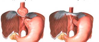
In adults, there are direct and oblique umbilical hernias. The formation of direct hernias is associated with thinning of the transverse fascia adjacent to the umbilical ring. In this case, the hernial sac enters the subcutaneous tissue directly through the umbilical ring. With oblique umbilical hernias, a hernial protrusion forms above or below the umbilical ring, passes through the gap between the linea alba and the transverse fascia (umbilical canal), then exits into the subcutaneous tissue through the umbilical ring.
According to the criterion of displacement, umbilical hernias are reducible and irreducible. A variant of an irreducible hernia is a strangulated umbilical hernia.
Types of hernias developing near the navel
The contents of the hernial sac most often become an intestinal loop, subcutaneous fatty tissue or greater omentum. Among diseases of the white line of the abdomen, epigastric and periumbilical hernia are often found. The latter has two main varieties:
- Direct - it is characterized by the formation of the transverse fascia, and the contents of the hernial formation exit through the umbilical ring.
- Oblique - this type is characterized by the presence of a double pouch (above and below the umbilical ring).
The patient can feel the peri-umbilical hernia formation on his own, but, unlike the umbilical one, this protruding “bag” does not reduce, disappear or become smaller, even if the patient takes a supine position.
Symptoms of an umbilical hernia
Fetal umbilical hernias usually occur with severe combined defects that are incompatible with life. With this type of defect, there is actually underdevelopment of the anterior abdominal wall, through which the hernial sac containing internal organs (intestines, liver) emerges. With embryonic umbilical hernias, there is often a splitting of the sternum, underdevelopment of the pubic joint, defects of the diaphragm, ectopia of the heart, and ectopia of the bladder. The death of a child, as a rule, occurs in the third year of life from peritonitis, pneumonia, sepsis; cases of a favorable outcome are rare.
Acquired umbilical hernias progress much more favorably. An umbilical hernia in a child is largely a cosmetic defect and is not accompanied by extensive symptoms and is not prone to strangulation. The bulge usually measures from 1 to 5 cm in diameter and becomes most noticeable when the child cries, coughs, or strains. At rest or in the supine position, the umbilical hernia is almost invisible.
An early symptom of an umbilical hernia is the appearance of a small spherical protrusion in the area of the umbilical ring. At first, this bulge is completely painless and can be easily reduced by pressing on the hernial protrusion. As adhesions form between the anterior abdominal wall and the hernial sac, the protrusion becomes irreducible. The severity of the symptoms of an umbilical hernia depends on its size, the size of the hernial orifice, the presence of adhesions, etc. With a narrow hernial orifice, prolapse of the hernial sac is accompanied by discomfort and abdominal pain, nausea, and chronic constipation.
Umbilical hernias can be complicated by strangulation, inflammation of the elements of the hernial sac, and coprostasis. Incarceration of an umbilical hernia is accompanied by sudden sharp pain, severe nausea, vomiting, the appearance of blood in the stool, delayed passage of gases and defecation, non-reducibility of the hernia in a horizontal position, and tension in the hernial protrusion.
Clinical picture in men and women
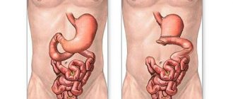
Protrusion of intestinal loops through the umbilical ring in adults manifests itself in different ways. Depending on the size of the hole, even the stomach can get into it, and a person’s well-being can sharply deteriorate. The umbilical ring is called the hernial orifice. This is the only place that is not covered by muscle tissue.
The organs that are hernial contents are located in a sac of peritoneum - thin connective tissue. In appearance, it is a spherical formation that protrudes beyond the anterior abdominal wall.
At the beginning of its formation, the hernia is small in size and is usually not visible in a horizontal position. The bulge increases over time and is more noticeable in an upright position when abdominal pressure increases.
Women over 40 years of age have a higher risk of developing an umbilical hernia than men. This is due to physiological characteristics - pregnancy, childbirth. Treatment of the disease is possible only surgically, since the hernial orifice will expand more and more over time.
Symptoms of umbilical hernia in adults
If it is impossible to set the hernial sac back, a person begins to have digestive problems - vomiting, nausea, impaired peristalsis. If the bladder gets into the hernial sac, problems with urination occur.
Pain from a navel hernia appears after physical exertion, prolonged coughing or constipation. When you feel the umbilical zone with your fingers, you can determine some empty cavity under the skin - a wide hernial orifice. Sometimes the patient may hear clicking sounds inside the abdomen.
With infringement, a completely different clinical picture is observed. The patient begins to experience severe pain, which can cause shock. The protrusion increases in size, the local body temperature in the area of the hernia rises, due to intestinal obstruction the muscles become tense, and the infringement becomes hard to the touch. Up to a certain point, the temperature rises, then begins to fall. This occurs due to necrotic processes in the intestines and acute intoxication of the body. If the patient is not operated on in time, death is possible, as with peritonitis. Once irreversible changes in tissues begin, a person’s chances of survival are very low.
Diagnosis of umbilical hernia
An examination for an umbilical hernia is carried out by a pediatrician or surgeon. When examining the patient, attention is drawn to the presence of a spherical protrusion in the umbilical region. Sometimes in the area of the hernia, the contours of intestinal loops and peristalsis are visible through thin skin. Palpation of the umbilical ring reveals a defect in the abdominal wall, a hernial sac, which usually includes a loop of intestine and a greater omentum. When the child cries and the abdomen tenses, the hernial protrusion increases.
Carrying out an endoscopic (EGD) and x-ray examination (herniography, x-ray of the stomach, x-ray of the passage of barium through the small intestine) allows you to get an idea of the contents of the hernial sac, assess the patency of the intestine and the severity of the adhesive process. The necessary information is clarified when performing an ultrasound of the abdominal organs and hernial protrusion.
Differential diagnosis of an umbilical hernia is carried out with a hernia of the white line of the abdomen, metastases to the navel of stomach cancer, and extragenital endometriosis of the navel.
Diagnostics
Usually the pathology is clearly visible visually. The doctor’s task at the first stage of diagnosis is to assess the contents of the hernial sac. For this purpose, the X-ray method and examination with an endoscope are used. X-rays are contrasted using barium. If the presence of adhesions is determined, there is a danger of imminent strangulation.
Ultrasound is used to clarify endoscopy and x-ray data. At this stage, it is important to differentiate an umbilical hernia from possible metastases of a stomach cancer to the intestine, a hernia of the white line and endometriosis of the umbilicus.
Gastroduodenoscopy is performed to determine the presence of inflammatory processes in the gastrointestinal tract, which complicate the course of the disease and affect the course of treatment.
Family history is of great importance. If close relatives had a similar problem, even in the initial stage it makes no sense to treat the disease with conservative methods - it will still manifest itself and threaten the person’s life.
Treatment of umbilical hernia
In children, an umbilical hernia can spontaneously regress, which is associated with physiological strengthening of the abdominal muscles. Therefore, up to 5 years of age, monitoring of umbilical hernia is indicated. During this period, it is recommended that children be placed on their tummy, a tonic massage of the anterior abdominal wall, general massage, exercise therapy, and the application of a therapeutic adhesive bandage to the navel are prescribed.
Surgical treatment (hernioplasty) is indicated for adults and children over 5 years of age with a non-regressed umbilical hernia. For umbilical hernia, two types of operations are used - hernioplasty with local tissues and using mesh implants.
With traditional plastic surgery, an incision is made in the infraumbilical (subumbilical) fold, the hernial sac is isolated and opened, its contents are reduced into the abdominal cavity, and the peritoneum is sutured. Then a duplication of the aponeurosis is formed in the transverse or vertical direction. In obese patients with excess skin-fat apron, abdominoplasty with navel transfer can be performed at the same time. The disadvantages of umbilical hernia hernioplasty with local tissues are the need for long-term (up to 1 year) restriction of physical activity and a high likelihood of relapse.
Hernioplasty with the installation of a mesh prosthesis does not have the disadvantages of the first operation. In this case, the mesh system can be installed under the skin above the aponeurosis (for large hernial orifices) or under the aponeurosis under the umbilical ring. This type of surgery reduces rehabilitation to 1 month, while the number of recurrences of umbilical hernia does not exceed 1%.
Treatment of umbilical hernia in adults
Treatment of umbilical hernia in adults is only surgical. Different types of operations are used, depending on the size of the hernia and the condition of the anterior abdominal wall.
Usually, surgery for an umbilical hernia, if there is no strangulation, is carried out as planned. During the first appointment, the doctor examines the patient, prescribes a preoperative examination and a date for hospitalization.
Preoperative examination in a patient with an umbilical hernia
Types of operations for umbilical hernia
An operation aimed at eliminating a hernial protrusion is called hernioplasty. Types of hernioplasty that are performed for umbilical hernia:
- Tension . The patient's umbilical ring is strengthened with his own tissue. In order to close the defect, they are pulled, which is why the operation got its name.
- Non-tensioned . To strengthen the umbilical ring, special synthetic mesh is used.
- Laparoscopic . The operation is performed without an incision, through punctures in the abdominal wall.
It is advisable to perform surgical intervention as early as possible, while the hernia is still small and can be reduced.
In adults, general anesthesia or local anesthesia can be used - injecting the navel area with anesthetic solutions. Tension hernioplasty
- The surgeon makes an incision and provides access to the hernial sac.
- Depending on the size of the hernial sac, it is either simply immersed in the stomach, or stitched and cut off.
- The umbilical ring is stitched and reinforced with adjacent tissues.
The disadvantage of this method is the high probability of relapse: after surgery, an umbilical hernia occurs again in 4-20% of patients.
Rehabilitation can last up to a year. Tension-free hernioplasty
The operation is performed in a similar way, but the surgeon uses a special synthetic mesh to strengthen the navel. Subsequently, it grows into surrounding tissues.
The advantage of tension-free hernioplasty is the relatively low likelihood of relapse.
The hernia occurs again on average in only 2 patients out of 100. The rehabilitation period lasts only 30 days, even for those people who play sports professionally. Laparoscopic hernioplasty
During laparoscopic surgery, a mesh implant is also used; it is installed through a puncture in the abdominal wall. The surgeon does not make a large incision, which significantly reduces postoperative rehabilitation time.
But there are also certain difficulties. Laparoscopic hernioplasty requires special equipment and trained surgeons. Not every hospital has this opportunity. Surgeries through a puncture are contraindicated in patients with pathologies of the respiratory and cardiovascular systems, with a large expansion of the umbilical ring.
Surgery for strangulated umbilical hernia
If an umbilical hernia is strangulated, surgery must be performed as an emergency.
The risk of strangulation does not depend on the size of the hernia - it increases the more the longer the patient does not see a doctor.
During surgery, the doctor opens the hernial sac and examines the organ that is inside. If it is not changed, then it is simply immersed in the stomach. If part of the organ is dead, it is excised. And if the doctor has doubts, he covers the organ with napkins soaked in warm saline and injects a solution of novocaine.
Rehabilitation after surgery for umbilical hernia in adults
- Usually, if the operation goes without complications, the patient is allowed to get up on the first day.
- In the postoperative period, wearing a special bandage is recommended (about a month when using mesh implants).
- On days 10-14, you can start doing therapeutic exercises, but you are prohibited from performing abdominal exercises.
- After the operation, daily dressings are performed, the sutures are removed on the 7th day (if they do not dissolve on their own).
- For pain, painkillers are prescribed.
- The doctor may also prescribe antibiotics, vitamins, and immunomodulators.
Wearing a bandage for an umbilical hernia
The bandage is not a treatment for an umbilical hernia.
It only helps, while wearing it, to correct the hernia and prevent it from being strangulated. Indications for wearing a bandage :
- After surgery for an umbilical hernia and in general during any surgical intervention when the incision passed through the navel.
- If there are temporary contraindications to surgery: acute diseases, exacerbations of chronic ones. After normalization of the patient's condition, surgical treatment is performed.
- Severe diseases: significant dysfunction of the cardiovascular and respiratory systems, exhaustion, old age, malignant neoplasms, etc.
- Late-stage pregnancy is also a contraindication for surgery.
The bandage is a wide belt made of elastic fabric, on the inner surface of which a special anatomically shaped pad is attached. She presses the navel and does not allow the hernia to protrude outward. The pelot can be connected to the bandage or attached to it with Velcro.
Traditional methods of treating umbilical hernia
An umbilical hernia in an adult is a disease that can only be eliminated with surgery.
“Spells” and gluing coins to the navel, methods that traditional medicine often recommends, “help” only small children, since their umbilical hernia can close on its own before the age of 5. This doesn't happen in adults.
Decoctions, infusions, and lotions with medicinal plants are ineffective. With their help, an umbilical hernia in an adult cannot be eliminated.
Forecast and prevention of umbilical hernia
For children, surgery for an umbilical hernia is easily tolerated; as a rule, it is not complicated by relapses and allows one to achieve a good cosmetic effect. If left untreated, an umbilical hernia can take a complicated course - become unreducible, strangulated, etc.
Measures to prevent umbilical hernia include: prevention of situations associated with straining of an infant (excessive screaming and crying, bloating, constipation, etc.), rational feeding, preventive massage and gymnastics aimed at strengthening the abdominal wall, treatment of intestinal dysbiosis, wearing prenatal bandage
Umbilical hernia in adults. Causes, symptoms, diagnosis and treatment
The umbilical ring is the weakest point on the anterior abdominal wall. Therefore, it is one of those places where hernial protrusions most often form. Loops of intestine, omentum and other organs can exit through the umbilical ring in adults.
Facts about umbilical hernias:
- constitute 5% of all abdominal hernias in adults;
- most often found in women over 40 years of age;
- the disease was first described by the ancient Roman physician Celsus, who lived in the 1st century AD;
- The first successful operation for an umbilical hernia was performed in France in 1885.