In this article we will look at how to treat lymphofollicular hyperplasia.
This is a pathological process in which cells grow uncontrollably. The process of proliferation of follicular tissue, forming the mucous and submucosal layers. This disease occurs in patients of any age and does not depend on their gender, food preferences, or place of residence.
Description
Lymphofollicular hyperplasia is diagnosed in the endocrine system, but most often the pathology affects the gastrointestinal tract. The predominance of the disease in the gastrointestinal tract is due to the presence of a large number of predisposing factors - a high level of stress, a large number of carcinogens, chronic pathologies of the gastrointestinal tract. In endocrine organs, hyperplastic changes develop against the background of systemic or endocrine disorders. For example, hyperplasia can be detected in the thymus gland if the patient has already been diagnosed with any pathology of the pituitary gland.
Reasons for development
The development of pathology is caused by a variety of negative effects of external and internal factors that lead to cell growth. Thus, lymphofollicular hyperplasia can occur against the background of concomitant problems - hyperglycemia, functional liver disorders, obesity. Scientists also consider hereditary predisposition to be a risk factor.
Pathology can develop for the following reasons:
- Impaired motility of the duodenum and stomach.
- Herpes virus infection.
- Immunity disorders.
- Constant stress, nervous disorders.
- Exposure to Helicobacter pylori.
- The presence of atrophic, autoimmune, chronic pathologies in the gastrointestinal tract (for example, gastritis in these forms).
- Blastomogenic effect.
- The influence of products that have specific tissue breakdown.
- Disruptions in the activity of the nervous regulation of the gastrointestinal tract.
- Hormonal abnormalities.
- Dysfunction of the internal secretion of the gastrointestinal mucosa.
Types of pathology
The pathology may be of the glandular type.
In medical practice, there are several characteristic types of lymphofollicular hyperplasia, which differ only in the characteristics of their course. The following types are distinguished:
- Focal appearance. It is an early form of the development of polyps, characteristic of certain areas of the mucosa. It appears as a slight growth with a changed structure. Upon careful examination, both single and multiple outgrowths are determined.
- Lymphoid. A significant increase in the number of lymphocytes, which pathologically change the structure of tissues. It occurs as a result of the penetration of viruses into the blood, provoking a reaction from the immune system.
- Lymphofollicular hyperplasia. Consequences of the negative influence of factors on flora and soft tissues, leading to cell proliferation.
- Hyperplasia of the integumentary epithelium of the stomach. A dangerous pathology that leads to the formation of tumors. This is due to the proliferation of the epithelium, which gradually changes its structure abnormally.
- Hyperplasia of the antrum of the stomach. Damage to the section that closes the stomach and serves to release food into the intestines. Often affects the duodenal bulbs.
- Glandular. The formation of polyp-shaped growths consisting of glandular cells.
- Polypoid. Benign neoplasm, single or multiple compactions with dense structural changes.
Symptoms
The symptoms of lymphofollicular hyperplasia strongly depend on the location of the pathology. What does this mean?
Its generalized signs include a decrease in albumin levels and an increase in the number of T-lymphocytes. There is a feeling of weakness and fever. It is important to note that if lymphofollicular hyperplasia is benign, then there are usually no symptoms. Negative symptoms are noted if the hyperplastic lesion of the gastrointestinal tract has a special course or is advanced. In this case, dyspepsia and epigastric pain often develop.
First symptoms of the disease
The disease is considered latent, so manifestation does not always occur in the first stages of formation. This significantly complicates the diagnosis of the disease and its presence is determined at the advanced stage. Common signs of pathology are fever, weakness and apathy, a quantitative increase in lymphocytes and a decrease in albumin levels. Benign tumors have no symptoms; malignant tumors are characterized by severe abdominal pain and dyspeptic disorders. Patients with lymphofollicular hyperplasia often suffer from nausea, heartburn and vomiting.
Stages
By stage, hyperplasia is classified according to the distribution and size of the follicles:
- At stage zero, lymphoid follicles are completely absent or weakly expressed, are randomly located, and are small in size.
- At the first stage, a single, diffuse proliferation of small follicles is observed.
- At the second stage, the follicles spread diffusely, densely, but do not unite into conglomerates.
- At the third stage, curling of the follicles is observed, sometimes in a colony of considerable size. The mucous membrane of the follicles is sometimes hyperemic.
- At the fourth stage, erosive areas are identified, pronounced hyperemia of the mucous membranes, on which fibrin plaque is present, is noted. The mucous membranes, in addition, acquire a matte color, and their vascular pattern intensifies.
Taking into account these features of the course and formation of lymphofollicular hyperplasia, some conclusions can be drawn:
- Clinical manifestations develop only at the 3rd-4th stage of the disease, when the patient develops pain in the abdominal region and intestinal bleeding appears.
- The disease can be detected at other stages only by chance, during the diagnosis of some other disorder. This is due to the absence of specific symptoms.
We will consider gastric hyperplasia below.
Lymphofollicular hyperplasia: causes, symptoms, diagnosis, treatment
In this article we will look at how to treat lymphofollicular hyperplasia.
This is a pathological process in which cells grow uncontrollably. The process of proliferation of follicular tissue, forming the mucous and submucosal layers. This disease occurs in patients of any age and does not depend on their gender, food preferences, or place of residence.
Description
Lymphofollicular hyperplasia is diagnosed in the endocrine system, but most often the pathology affects the gastrointestinal tract.
The predominance of the disease in the gastrointestinal tract is due to the presence of a large number of predisposing factors - a high level of stress, a large number of carcinogens, chronic pathologies of the gastrointestinal tract.
In endocrine organs, hyperplastic changes develop against the background of systemic or endocrine disorders. For example, hyperplasia can be detected in the thymus gland if the patient has already been diagnosed with any pathology of the pituitary gland.
Reasons for development
The development of pathology is caused by a variety of negative effects of external and internal factors that lead to cell growth. Thus, lymphofollicular hyperplasia can occur against the background of concomitant problems - hyperglycemia, functional liver disorders, obesity. Scientists also consider hereditary predisposition to be a risk factor.
Pathology can develop for the following reasons:
- Impaired motility of the duodenum and stomach.
- Herpes virus infection.
- Immunity disorders.
- Constant stress, nervous disorders.
- Exposure to Helicobacter pylori.
- The presence of atrophic, autoimmune, chronic pathologies in the gastrointestinal tract (for example, gastritis in these forms).
- Blastomogenic effect.
- The influence of products that have specific tissue breakdown.
- Disruptions in the activity of the nervous regulation of the gastrointestinal tract.
- Hormonal abnormalities.
- Dysfunction of the internal secretion of the gastrointestinal mucosa.
Symptoms
The symptoms of lymphofollicular hyperplasia strongly depend on the location of the pathology. What does this mean?
Its generalized signs include a decrease in albumin levels and an increase in the number of T-lymphocytes. There is a feeling of weakness and fever.
It is important to note that if lymphofollicular hyperplasia is benign, then there are usually no symptoms.
Negative symptoms are noted if the hyperplastic lesion of the gastrointestinal tract has a special course or is advanced. In this case, dyspepsia and epigastric pain often develop.
Stages
By stage, hyperplasia is classified according to the distribution and size of the follicles:
- At stage zero, lymphoid follicles are completely absent or weakly expressed, are randomly located, and are small in size.
- At the first stage, a single, diffuse proliferation of small follicles is observed.
- At the second stage, the follicles spread diffusely, densely, but do not unite into conglomerates.
- At the third stage, curling of the follicles is observed, sometimes in a colony of considerable size. The mucous membrane of the follicles is sometimes hyperemic.
- At the fourth stage, erosive areas are identified, pronounced hyperemia of the mucous membranes, on which fibrin plaque is present, is noted. The mucous membranes, in addition, acquire a matte color, and their vascular pattern intensifies.
Taking into account these features of the course and formation of lymphofollicular hyperplasia, some conclusions can be drawn:
- Clinical manifestations develop only at the 3rd-4th stage of the disease, when the patient develops pain in the abdominal region and intestinal bleeding appears.
- The disease can be detected at other stages only by chance, during the diagnosis of some other disorder. This is due to the absence of specific symptoms.
We will consider gastric hyperplasia below.
Hyperplasia affecting the gastric mucosa
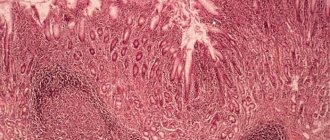
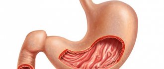
The gastric mucosa has a very complex structure, which is due to the performance of many functions, including protective and secretory. In addition, it takes part in the process of peristalsis.
Lymphofollicular hyperplasia of the gastric mucosa is the process of excessive proliferation of epithelial cells with simultaneous thickening of the wall of the mucous membranes. Very often the pathology is accompanied by the appearance of polyps and growths. The reasons for the development of gastric hyperplasia are usually attributed to hormonal changes and neurological disruptions.
Hyperplasia very rarely transforms into oncology. In most cases, the appearance of cancer cells is facilitated by epithelial dysplasia, when the cells that form the mucosa are transformed into cells with a pronounced atypical structure.
The most dangerous disease is mucosal metaplasia, which is characterized by the development of digestive dysfunction and a high risk of developing malignant tumors.
The main tasks of a gastroenterologist for lymphofollicular hyperplasia are making a diagnosis and prescribing the correct treatment. Moreover, treatment methods must be selected individually.
How does gastritis with lymphofollicular hyperplasia occur?
Pathology affecting the antrum of the stomach
Statistical data show that such hyperplasia in the antrum of the stomach develops not only in the presence of chronic gastritis, which is provoked by exposure to Helicobacter pylori, but also against a background of weakened immunity. Immune changes together with gastritis are diagnosed, as clinical practice shows, in conditions of low acidity, which, in turn, is a prerequisite for the appearance of autoimmune pathologies.
In childhood
A study of cases of the development of the disease in childhood made it possible to determine that in the antrum lymphofollicular hyperplasia develops as a result of autoimmune rheumatic pathologies, and not the activity of bacteria. Undoubtedly, the presence of pathogenic microflora in combination with autoimmune abnormalities significantly increases the likelihood of the disease occurring.
Very often, changes in the mucous membranes cause the development of polyps localized in the antrum. Polyps are inflammatory in nature and occur in 70-90% of cases. Externally they look like dense formations with a rounded cylindrical shape, a wide base and a flat top.
Lymphofollicular hyperplasia of the ileum
The ileum is the lower part of the small intestine. The inside is lined with mucous membrane, on which there are many villi. On its surface there are also capillaries and lymphatic vessels that take part in the absorption of useful substances.
In the ileum, lymphofollicular hyperplasia is formed as a result of proliferative processes in the intestinal wall and immunodeficiency. Clinically, the pathological condition is manifested by the following symptoms:
- Noticeable suppression of the immune system.
- Dramatic weight loss.
- Abdominal pain.
- Presence of blood and mucus in the stool.
- Loose stools, frequent urge to have bowel movements.
There is a change in the main indicators of the immune system: a significant increase in the percentage of T-lymphocytes.
Differentiation of the disease
Differentiation of the disease occurs on the basis of laboratory tests of stool, urine, blood and the results of fibrin fiber endoscopy. Most often, lymphofollicular dysplasia can be diagnosed when it affects the terminal zone of the ileum.
This suggests that the pathological process is secondary in nature and therapeutic action on it is not required. As a preventive and therapeutic measure, a strict diet may be recommended, in which a number of foods are prohibited.
In cases where the inflammation is serious and there is a suspicion of Crohn's disease or cancer, surgical intervention or drug therapy is indicated.
Hyperplastic changes in the lymph nodes are a clinical symptom, accompanied by excessive growth of lymph node cells and a gradual decrease in their number due to degeneration and changes in structure.
As a rule, lymph node hyperplasia is the body’s immune response to a variety of infections that have entered the body. Lymphadenitis can also be of bacterial, viral, or oncological origin.
Thus, submandibular lymphadenitis often develops against the background of tonsillitis, scarlet fever, felinosis, caries, diphtheria, mumps and other diseases.
Diagnostics
The disease is difficult to detect in the initial stages of its development, since it is practically asymptomatic. Quite often, lymphoid follicles are detected during colonileoscopy for other diseases.
Other diagnostic methods that allow one to examine an enlarged mucous layer in the intestines and stomach include: sigmoidoscopy, FGDS, colonoscopy, radiography using a contrast agent. Using X-rays, it is possible to assess the extent of the spread of pathological cells.
If lymphofollicular hyperplasia is detected, the patient is advised to undergo periodic examinations, due to the likelihood of abnormal areas degenerating into malignant tumors.
The treatment of the disease is carried out by gastroenterologists, surgical oncologists, and oncologists.
Therapy
In cases where lymphofollicular hyperplasia of the gastrointestinal tract occurs with the appearance of obvious signs of pathology, therapy aimed at reducing acidity in the stomach and suppressing the activity of Helicobacter pylori is indicated. Therapy involves the mandatory elimination of gastritis by following a diet and using medications, including antibiotics.
Treatment of lymphofollicular hyperplasia should be comprehensive.
In the presence of malignant tumors, surgical intervention is indicated. In case of hyperplasia in the digestive system, excision of the affected areas of the intestine and gastric resection are performed. The duration of the rehabilitation period depends on the nature and severity of the disease, the general condition of the patient and the success of the operation.
When identifying pathological foci of hyperplasia in the hematopoietic and endocrine systems that have signs of a malignant process, combination therapy is required, which combines chemotherapy and surgical techniques.
Therapy for benign lymphofollicular hyperplasia is usually not required.
Source: https://FB.ru/article/405409/limfofollikulyarnaya-giperplaziya-prichinyi-simptomyi-diagnostika-lechenie
Hyperplasia affecting the gastric mucosa
The gastric mucosa has a very complex structure, which is due to the performance of many functions, including protective and secretory. In addition, it takes part in the process of peristalsis.
Lymphofollicular hyperplasia of the gastric mucosa is the process of excessive proliferation of epithelial cells with simultaneous thickening of the wall of the mucous membranes. Very often the pathology is accompanied by the appearance of polyps and growths. The reasons for the development of gastric hyperplasia are usually attributed to hormonal changes and neurological disruptions. Hyperplasia very rarely transforms into oncology. In most cases, the appearance of cancer cells is facilitated by epithelial dysplasia, when the cells that form the mucosa are transformed into cells with a pronounced atypical structure. The most dangerous disease is mucosal metaplasia, which is characterized by the development of digestive dysfunction and a high risk of developing malignant tumors.
The main tasks of a gastroenterologist for lymphofollicular hyperplasia are making a diagnosis and prescribing the correct treatment. Moreover, treatment methods must be selected individually.
How does gastritis with lymphofollicular hyperplasia occur?
What is lymphoid hyperplasia
The mucous membrane of the digestive tract, including the ileum, is susceptible to the development of a number of diseases. Lymphoid hyperplasia manifests itself in the form of rapid proliferation of mucosal cells.
As a result of the spread of the pathological process, follicular tissue begins to form. In medicine, local and diffuse forms are distinguished. In the first case, the growth has a benign course.
The diffuse form is characterized by the appearance of small follicles. They are malignant.
Pathology is established in adults and children. Experts have not established a connection between ileal disease and gender or age category. There was also no relationship with the disease and eating habits.
Scientists believe that the main cause of lymphoid hyperplasia of the ileum is endocrine disorders.
Pathology affecting the antrum of the stomach
Statistical data show that such hyperplasia in the antrum of the stomach develops not only in the presence of chronic gastritis, which is provoked by exposure to Helicobacter pylori, but also against a background of weakened immunity. Immune changes together with gastritis are diagnosed, as clinical practice shows, in conditions of low acidity, which, in turn, is a prerequisite for the appearance of autoimmune pathologies.
Treatment
You should not try to cure the disease on your own; if you detect the first signals of an impending disease, you should consult a gastroenterologist for advice
Benign lymphofollicular hyperplasia does not require treatment.
If a malignant growth of gastric lymphoid tissue is diagnosed at an early stage, antibiotic therapy can help eliminate Helicobacter pylori.
Most lymphofollicular hyperplasia of the gastric antrum respond to modern treatment methods - radiotherapy and chemotherapy.
In later stages, surgery may help by removing only the affected part or the entire stomach. Complete removal of the stomach is called a gastrectomy.
Tumors that are limited to the inner layer of the stomach wall (mucosa) can be removed during gastroscopy. In this case, only part of the tumor and immediately adjacent tissue are removed. For deep-seated tumors, part or all of the stomach, including surrounding lymph nodes, the spleen, and part of the pancreas, must be removed. To restore the passage of food, the rest of the stomach, or the end of the esophagus, is connected to the small intestine.
Additional chemotherapy (given both before and after surgery) may improve the chances of survival for patients with locally advanced tumors that have an increased risk of recurrence.
If the tumor has spread to the abdomen (peritoneal carcinomatosis), the patient's life can be prolonged by surgical removal of the affected peritoneal membrane in combination with so-called hyperthermic intraperitoneal chemotherapy.
If the tumor cannot be completely removed, surgery is not performed. In this case, drug treatment (chemotherapy, possibly in combination with other medications) can relieve symptoms, prolong and improve quality of life.
If the stomach is severely compressed by a tumor, inserting a plastic or metal tube (called a stent) may help you eat normally.
Many patients suffer from digestive problems after surgery.
In childhood
A study of cases of the development of the disease in childhood made it possible to determine that in the antrum lymphofollicular hyperplasia develops as a result of autoimmune rheumatic pathologies, and not the activity of bacteria. Undoubtedly, the presence of pathogenic microflora in combination with autoimmune abnormalities significantly increases the likelihood of the disease occurring.
Very often, changes in the mucous membranes cause the development of polyps localized in the antrum. Polyps are inflammatory in nature and occur in 70-90% of cases. Externally they look like dense formations with a rounded cylindrical shape, a wide base and a flat top.
Causes and risk factors for the development of hyperplastic polyps
Polyps most often develop in men between 40 and 60 years of age. In 80% of cases, doctors find single polyps when examining patients. Group and multiple neoplasms are less common. In 5-6% of patients, proctologists detect diffuse colon polyposis.
The development of hyperplastic polyps is provoked by the following factors:
- The nature of the diet with a predominance of refined foods in the diet, which contribute to constipation and prolonged retention of contents in the large intestine;
- Colon dysbiosis, which reflects a violation of local and decreased general immunity, contributes to changes in the differentiation and restoration of mucosal cells;
- Concomitant diseases of the biliary system and disorders of the production of bile acids, which have a mutagenic effect on the mucous membrane of the large intestine.
Active chronic inflammation and dysplasia of the mucous membrane play a certain role in the development of hyperplastic polyps.
Lymphofollicular hyperplasia of the ileum
The ileum is the lower part of the small intestine. The inside is lined with mucous membrane, on which there are many villi. On its surface there are also capillaries and lymphatic vessels that take part in the absorption of useful substances.
In the ileum, lymphofollicular hyperplasia is formed as a result of proliferative processes in the intestinal wall and immunodeficiency. Clinically, the pathological condition is manifested by the following symptoms:
- Noticeable suppression of the immune system.
- Dramatic weight loss.
- Abdominal pain.
- Presence of blood and mucus in the stool.
- Loose stools, frequent urge to have bowel movements.
There is a change in the main indicators of the immune system: a significant increase in the percentage of T-lymphocytes.
Intestinal diseases treatment
Intestinal diseases. Treatment of intestinal diseases with folk remedies
Intestinal hyperplasia
● Intestinal hyperplasia is a slight thickening of the folds of the intestinal mucosa. Most patients with this intestinal disease have a question: “Is it possible to stop the growth of intestinal epithelium with signs of hyperplasia?” This is possible by following the recommendations below.
● First of all, you need to strengthen your immune system. You will learn how to achieve this by reading the article “Healing herbs to boost immunity .” By increasing the body's defenses and resistance, you gradually get rid of many diseases. Eat more unprocessed fresh vegetables, herbs, berries and fruits every day; in total up to 600 g per day. To rid your body of toxins, drink at least two liters of water, adding to your taste aromatic spices such as mint, cloves, cinnamon, cumin and dill, which enhance the taste. Consume up to 100 g of fermented milk bifidum-bacterin daily.
● And now I bring to your attention two effective traditional medicine recipes for the treatment of intestinal hyperplasia:
#8212; for 12 hours, infuse a teaspoon of mountain arnica herb in a glass of boiling water and drink ⅓ glass of infusion three times a day before meals for five minutes;
#8212; rinse the dandelion roots thoroughly, wilt them until the juice flow stops, then get the powder by grinding them in a coffee grinder; take half a teaspoon of powder three times a day, half an hour before meals, with a small amount of water. Treat with herbs for 4 to 6 weeks, 2-3 times a year.
● Intestinal diseases should also be treated with simple physical exercises: morning 10-minute exercises and dousing with cold water. Depending on your health, practice brisk walking or jogging for thirty minutes three times a week. Swimming once a week and cycling twice a week for one hour will help you.
Spastic colitis complicated by adenoma and hemorrhoids
● This diagnosis was given to one patient who complained of pain in the anus and mucus discharge from the rectum. More precisely, we are dealing here with distal proctitis - inflammation of the mucous membrane of the rectal outlet. In such cases, it is advisable to conduct an endoscopic examination of the colon and rectum and bacteriological examination of stool.
● First of all, if you have chronic intestinal colitis and proctitis, you should follow a special non-irritating diet. If you are constipated, eat foods with the addition of wheat bran; if you have loose stools (diarrhea), limit foods rich in fiber. Medicinal microenemas are effective:
#8212; first seven days: in the morning after bowel movement #8212; microenema with rotokan (half a glass of boiled water, a teaspoon of raw materials); at night - with 10 ml of vinylin;
#8212; second seven days: in the morning after bowel movement - microenema with 50 ml of protargol 0.5%; at night - with 10 ml of carotoline.
● Next, inject into the rectum for fourteen days in the morning after stool 0.5 cm of methyluracil ointment squeezed out of a tube with a methyluracil suppository; at night - insert a “Tykveol” candle into the anus.
● If you have dysbacteriosis, take one tablespoon in the morning after breakfast - normofolin-L, and before dinner - normofolin-B. Treat for 10 days. Antispasmodics will help you get rid of pain: buscopan or mebeverine - one tablet three times a day for three weeks.
Trophic ulcers near the anus with copious discharge
● Spicy foods with red pepper, horseradish or mustard, as well as alcohol abuse, can provoke excessive mucus secretion. Benign villous tumors of the sigmoid and rectum can also cause mucus formation. X-ray, endoscopic and other examinations (colonoscopy) will help clarify the diagnosis.
● The villous tumor is removed in the hospital using electrocoagulation or excision of the affected area of the intestine. As a rule, the prognosis is favorable. If there are contraindications to surgery, use herbal medicine:
#8212; let a tablespoon of celandine herb brew in 200 ml of boiling water for 15-20 minutes, then before going to bed, inject a cooled ¼ cup of infusion into the rectum for 10 days in a row. Then undergo a control examination of the intestines. As clinical trials have shown, after such treatment the tumor component disappears and its growth is inhibited;
#8212; Immunotherapy described above is an addition to the treatment of the disease.
● Grind the mistletoe herb and fill a 700 ml bottle with it, fill it with vodka and let it steep for three weeks; do not forget to periodically shake the contents of the bottle. Drink 30-40 drops of tincture three times a day before meals for half an hour for a year.
● The homeopathic remedy podophyllum will help you prevent the appearance of new formations in the intestines - 10 drops before meals with water three times a week, then a seven-day break; the course of treatment is six months.
● All patients are recommended to undergo endoscopic examination to clarify the diagnosis. Many patients refuse colonoscopy due to pain during this procedure. But pain is easily relieved by taking analgesics, anesthetics, or, as a last resort #8212; anesthesia. Colonoscopy helps the doctor assess the condition of the disease not by indirect signs, but by the results of accurate observations and promptly prescribe adequate treatment for the disease.
Atony and chronic constipation
● The reason for this may be dolichosigma - an elongated sigmoid colon, which gives kinks and slight inversion of excess loops.
● To normalize the motor-evacuation function of the large intestine and prevent its atony, take proserin thirty minutes before meals, 10-15 mg 2-3 times a day. Or dibazol 20-50 mg before meals two hours or two hours after meals, also 2-3 times a day. Duration of treatment is 2-3 weeks with 2-3 month breaks.
● Take vitamins at the same time: Complivit - 1 tablet twice a day for 3-4 weeks. For constipation, use Vaseline oil according to Art. spoon two hours after dinner. The course is thirty days.
Differentiation of the disease
Differentiation of the disease occurs on the basis of laboratory tests of stool, urine, blood and the results of fibrin fiber endoscopy. Most often, lymphofollicular dysplasia can be diagnosed when it affects the terminal zone of the ileum. This suggests that the pathological process is secondary in nature and therapeutic action on it is not required. As a preventive and therapeutic measure, a strict diet may be recommended, in which a number of foods are prohibited. In cases where the inflammation is serious and there is a suspicion of Crohn's disease or cancer, surgical intervention or drug therapy is indicated.
Symptoms of the disease
Symptoms of hyperplasia are quite varied and individual for each person.
The most common are the following:
- Increased body temperature.
- Pain in the stomach area.
- Weakness.
- Dyspeptic disorders - belching, heartburn, bad breath, feeling of nausea, gag reflexes, gas formation. These symptoms are similar to those of most gastrointestinal diseases and more often torment the patient after eating or, conversely, on an empty stomach. Disorders can occur at night.
It is impossible to independently make a correct diagnosis and identify lymphofollicular hyperplasia. Only a doctor, after taking the necessary measures, will be able to identify the disease and prescribe treatment.
Lymph node hyperplasia
Hyperplastic changes in the lymph nodes are a clinical symptom, accompanied by excessive growth of lymph node cells and a gradual decrease in their number due to degeneration and changes in structure. As a rule, lymph node hyperplasia is the body’s immune response to a variety of infections that have entered the body. Lymphadenitis can also be of bacterial, viral, or oncological origin. Thus, submandibular lymphadenitis often develops against the background of tonsillitis, scarlet fever, felinosis, caries, diphtheria, mumps and other diseases.
Types of colon polyps
Hyperplastic polyp of the colon - what does it mean? Hyperplastic polyps account for 28 to 42% of all colon polyps. Most often they are localized in the left half of the colon, mainly in the lower sections, are multiple in nature and have no clinical manifestations. During an endoscopic examination, proctologists in most cases identify flattened “sessile” formations measuring 0.1–0.5 cm, rarely 1.0 cm, with a pearl-colored surface.
There are three histological variants of hyperplastic polyps:
- Rich in goblet cells;
- Microvesicular;
- Poor mucin.
Most hyperplastic polyps show no signs of cellular atypia. The diameter of small hyperplastic polyps of the large intestine does not exceed 0.5 cm. They are of normal color, slightly rise above the level of the mucous membrane, and have a soft consistency. These formations are characterized by elongation and cystic expansion of the crypts.
In such polyps, the epithelium is saw-toothed and the number of goblet cells is reduced. Morphologically, not being true adenomas, miliary hyperplastic polyps during histological examination often reveal a picture of atypia and proliferation of the epithelium with an increase in the amount of chromatin in the nuclei and an abundance of cell division (mitoses).
Benign glandular polyps, or tubular adenomas, are pedunculated or sessile hyperplasia of the mucosa. These are true adenomatous polyps. Upon examination, they are close in structure to the surrounding mucous membrane of the large intestine, but have a denser consistency, move along with the mucous membrane, and rarely ulcerate or bleed.
Adenomatous polyps are represented by glands that are lined with homogeneous glandular epithelium. Their cystic expansion is often noted. According to the degree of morphological differentiation of the epithelium, three groups of tubular adenomas are distinguished: with mild, moderate and significant dysplasia. With electron microscopy, morphologists often detect changes in them that are typical of cancer cells.
Juvenile polyps are not hyperplastic neoplasms. They lack enlargement of glands and changes in the glandular epithelium. These formations are quite large, sometimes hanging into the intestinal lumen on a long stalk, smooth, bright red or cherry color.
Diagnostics
The disease is difficult to detect in the initial stages of its development, since it is practically asymptomatic. Quite often, lymphoid follicles are detected during colonileoscopy for other diseases.
Other diagnostic methods that allow one to examine an enlarged mucous layer in the intestines and stomach include: sigmoidoscopy, FGDS, colonoscopy, radiography using a contrast agent. Using X-rays, it is possible to assess the extent of the spread of pathological cells.
If lymphofollicular hyperplasia is detected, the patient is advised to undergo periodic examinations, due to the likelihood of abnormal areas degenerating into malignant tumors.
The treatment of the disease is carried out by gastroenterologists, surgical oncologists, and oncologists.
Early diagnosis is the basis of treatment
All diagnostic measures are carried out to establish the characteristics of the disease; it is impossible to diagnose the disease without the use of medical equipment. Treatment of lymphofollicular hyperplasia begins with diagnosis and examination of the patient. For this they widely use:
The FGDS procedure will help determine the presence of pathology.
- Radiography, which helps determine the contours, shape and size of polyps on the walls.
- Endoscopy. Carry out for histological analysis of polyp tissue.
- Fibrogastroduodenoscopy. Used for visual inspection of the gastrointestinal tract. The procedure is appropriate for making a diagnosis and determining the nature of the formation: polyp or tumor.