Location
The stomach is located in the upper abdominal cavity. Human anatomy reliably hides the organ under the cover of the lower ribs and thus protects it from mechanical damage.
In front it is adjacent to the abdominal wall, left hypochondrium, left lung, diaphragm and liver, and behind it is adjacent to the lesser omentum, diaphragm, spleen, left adrenal gland, upper part of the left kidney, splenic artery, pancreas and transverse colon.
The stomach is fixed at both ends, but is mobile between them, constantly changing shape depending on its filling.
Not a diagnosis, but a condition
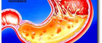
Cascade stomach is a term used by radiologists and doctors working in endoscopy rooms where EGD is performed. The stomach is a mobile organ, capable of functionally changing its shape depending on the patient’s posture, intake of food and drink, and other factors.
If a person is hungry, then during endoscopy or x-ray the doctor sees the organ in the form of a flat empty stocking, resembling a horn, with almost touching walls. When a contrast agent (barium) enters inside, the shape of the organ changes and takes on the appearance of a hook. These shape changes are clearly visible in photo 1.
Photo 1
If a person's stomach is filled with food or liquid, it is able to stretch, flatten, and its shape will resemble a giant sausage.
In the presence of a cascade inflection, the shape of the stomach is voluminous and convex in the area of the cardia, fundus and body, and narrowed in the area of the antrum and pylorus.
Photo 2 shows a cascading stomach, look at it carefully.
photo 2
The X-ray image clearly shows the bending of the organ around its own axis with the formation of two stages of the cascade - the upper wide and the lower narrow.
Such variability can be an innate structural feature or acquired.
According to radiologists, a hook-shaped stomach occurs in 60–80%, a horn-shaped stomach in 20%, and a stocking-like stomach in 5–9% of cases.
The cascade form accounts for up to 5% of survey results , which indicates a rare form.
Structure
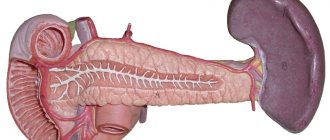
The organ can be called part of the digestive tract chain and, undoubtedly, its most important link. It is located in front of the duodenum and, in fact, is a continuation of the esophagus. The stomach and the anatomy of the tissues lining its walls are composed of: mucous, submucosal, muscular and serous membranes.
The mucous membrane is the place where acid is produced and secreted.
The submucosa is a layer composed of connective tissue that separates the mucosa from the muscular outer surface.
Muscle - consists of fibers, which are divided into several types, named according to their location in the organ. These are the inner oblique layer, the middle circulation layer and the outer longitudinal layer. All of them are involved in uniform mixing and grinding of food, as well as its further movement along the tract.
The final layer, serosa, is a connective tissue that lines the outer walls of the stomach and prevents it from sticking to neighboring organs.
Behind the organ is the pancreas and the greater omentum. The main areas of the stomach structure and anatomy consist of: the esophageal sphincter (cardiac sphincter), fundus, body, antrum (pyloric) and pylorus. In addition, it has a major curvature (posterior convex part) and a lesser curvature (anterior concave part), which are located on the left and right sides, respectively. The esophageal sphincter is located in the cardiac region and controls the flow of material into the stomach. The bottom is its upper section, the wall of which is formed by the superior curvature, and the body represents the main area of the organ. The final part is the antrum, serves as the exit and entrance to the small intestine and ends with the pyloric sphincter (pylorus).
Secretory
One of the main tasks of the stomach is to produce gastric juice. Gastric juice contains hydrochloric acid (on average up to 0.5%), water, mineral salts and enzymes, the main of which is pepsin, which is responsible for the breakdown of proteins.
Special glands are responsible for the production and secretion of various substances; there are three types of glands in the gastric mucosa: main, parietal and accessory. Most of all, on the mucous membrane of the main glands, they produce non-protein substances - pepsinogens, from which the enzyme pepsin is then formed. The parietal glands are involved in the synthesis of hydrochloric acid, which, in turn, is responsible for the “disinfection” of the stomach and helps break down food faster. And finally, accessory glands: they are involved in the production of special mucus that covers the inner wall of the stomach and protects it from the effects of hydrochloric acid and enzymes.
Article on the topic
Get over it. The most common questions about the functioning of the stomach
Holes
Cardiac opening. Lies near the heart where the esophagus enters the gastric mass. This opening does not have anatomical constipation, but contains a special mechanism through which food does not flow back. In this system, the lower circular smooth muscle fibers of the esophagus serve as a physiological sphincter.
Pyloric opening. Formed by the pyloric canal, which attaches to the first part of the small intestine - the duodenum - and creates an exit route for chyme. It differs from the cardiac one in that it has a pyloric sphincter with a valve. It consists of a circular muscular membrane that thickens around it. The pylorus controls the rate of release of stomach contents into the duodenum.
Structure of the stomach
In a healthy person, the muscular one is located on the left side of the body, has 4 sections, three of which enter the digestive sac, and the fourth serves as an evacuator for food residues.
- Cardiac (lat. pars cardiaca) department. The muscular part of the junction of the esophagus with the stomach, which is located in close proximity to the heart and does not allow food to be thrown back into the esophagus.
- Fundus (bottom, Latin fornix ventricul) or vault . A dome-shaped area located above the cardiac region, near the ribs and producing gastric juice with the help of glands. Also promotes the release of trapped air with food.
- Body of the stomach (lat. corpus ventriculi). The most voluminous part of the organ, intended for collecting and digesting food, occupies 2/3 of the total size and is located between the bottom of the stomach and the pylorus.
- Pyloric department (lat. pars pyloric) or pylorus . The muscular part of the stomach, together with the connection with the duodenum, has parts - a canal and a cave, designed to move food through the intestines. Located in close proximity to the spine.
The organ grows and changes during a person’s life, acquiring an individual shape. It depends on lifestyle, pathological processes and injuries/surgical interventions. In people who overeat, its shape is baggier, shifted to the center of the abdomen.
Two curvatures
Less curvature. It is also part of the anatomy of the stomach, or rather, its right outer border, and stretches from the cardiac opening to the pylorus. It faces the liver and is in contact with it and other organs.
Great curvature. Significantly longer than the smaller one and runs to the left of the cardial opening, along the bottom and left border of the stomach. Its anatomy extends to the pylorus; The hepatogastric ligament of the lesser omentum diverges from the upper part, and the greater omentum diverges from the lower part.
Sections of the stomach
- Bottom. The dome-shaped upper part, which projects up and to the left of the cardiac opening. Usually it fills with an excess of gases and releases them back through the esophagus as a belch.
- Body. Located between the cardiac and antral sections.
- Pyloric department. Continues the body and anatomy of the stomach, being at the very bottom of the organ and ending with the pylorus.
- Mucous. A thick and vascular surface with numerous folds, known as wrinkles, which have a predominantly longitudinal direction. When filled with food, these folds are flattened, expanding the boundaries of the organ. They contain glands and gastric pits
Stomach walls
The walls consist of muscle tissue and contain three layers: longitudinal, round and oblique.
Longitudinal. The most superficial fibers of the muscle wall, concentrated along the curvature.
Circular. Lies under the longitudinal ones and surrounds the body of the stomach. It thickens significantly at the pylorus to form the sphincter. Only a few circular fibers were found in the bottom area.
Oblique. Forms the innermost lining of the stomach. The anatomy of this muscle tissue is arranged as follows: it loops along the bottom and runs along its anterior and posterior walls, running almost parallel to the lesser curvature.
Blood supply to the stomach
The stomach receives an extensive blood supply.
Left artery. It arises directly from the celiac trunk and supplies blood, according to its name, to the left side of the stomach, partially to its right side, as well as the esophagus.
Right artery. It is a continuation of the hepatic artery and stretches from the upper border of the pylorus to the lesser curvature. Then it diverges along the lower right side of the stomach and at the end merges with the left gastric artery and anatomy. You can see a photo of the blood supply diagram of the entire organ below.
Short arteries. These are small branches that diverge from the large splenic artery, supply the lower part of the organ and connect with the left and gastroepiploic arteries.
Left gastroepiploic artery. It also continues the splenic artery, passes along the greater curvature and between the layers of the greater omentum.
Right gastroepiploic artery. A branch of the gastroduodenal artery that moves to the left and joins the left gastric artery. It diverges along the right side of the organ and the upper part of the duodenum.
The stomach has exactly the same number of veins as arteries, and they are called exactly the same. The right and left drain directly into the portal vein. The short and left gastroepiploic vein drain into the splenic vein, and the right gastroepiploic vein drains into the superior mesenteric vein.
Innervation
The stomach receives signals from the sympathetic and parasympathetic nervous systems. Sympathetic fibers are derived from the celiac plexus, and parasympathetic fibers are derived from the right and left vagus nerves.
The vagus nerves in the chest form the anterior and posterior vagal trunks. The anterior trunk is formed predominantly by the left nerve. It enters the abdominal cavity along the outer surface of the esophagus and stretches along the anterior edge of the stomach. The posterior nerve, on the contrary, is located along the posterior walls of the organs.
The pyloric sphincter receives motor fibers from the sympathetic system and inhibitory fibers from the parasympathetic system.
Functions
The main tasks in the anatomy of the stomach include killing bacteria, processing food, and then pushing it further into the small intestine while maintaining a constant rate of release of material.
The pH inside the organ is maintained at a very high acidic level, which helps digestive enzymes such as pepsin break down food so it can move along the tract. Finally, the stomach, along with the small intestine, takes part in the absorption of vitamins.
After chewing and swallowing food, it moves down the esophagus, then entering the stomach. There it remains for a certain amount of time (depending on the nature of the food) until it reaches the appropriate consistency for digestion and absorption in the small intestine. The organ mixes food with its secretions, forming a semi-liquid paste.
Thus, after the chemical and mechanical breakdown of food, the stomach controls the amount of mass that passes further. This is to ensure that food is not passed forward faster than it is processed.
Motor function
Further digestion of food occurs in the stomach. The motor function of the stomach is the accumulation of food mass, its mechanical processing and further movement into the intestine.
During meals and in the first minutes after this, the stomach is relaxed, which promotes the accumulation of food in it and ensures the release of secretions. Next, contractile movements begin, which are provided by the muscle layer. In this case, the food mass is mixed with gastric juice.
The muscles of the organ are characterized by the following types of movements:
- Peristaltic (wavy).
- Systolic - occur in the pyloric region.
- Tonic - help reduce the size of the stomach cavity (its bottom and body).
After eating, peristaltic waves are weak at first. By the end of the first hour after a meal, they intensify, which promotes the movement of the food bolus towards the exit from the stomach. The pressure in the pyloric region of the stomach increases. The pyloric sphincter opens and part of the food mass enters the duodenum. The remaining majority of this mass is returned to the pyloric region. The evacuation function of the stomach is inseparable from the motor function. They ensure grinding and homogenization of the food mass and thereby contribute to better absorption of nutrients in the intestines.
Sphincters
They are circular muscles associated with the stomach, structure and function. The anatomy of these organs opens and closes passages for food to enter and exit.
Thus, the first check valve (cardiac) is located between the esophagus and the stomach, allowing food to pass and helping to prevent food from being retained in the esophagus. If the sphincter does not work properly, acid flows back and causes what is commonly known as heartburn.
Another valve (the pylorus) allows food to pass from the stomach to the small intestine. As mentioned above, this sphincter helps the stomach control how much food is sent into the duodenum at one time.
MAIN FUNCTIONS AND TASKS OF THE GASTROINTESTINAL TRACT
The main tasks of the gastrointestinal tract are mechanical and chemical processing of food, absorption of nutrients (including from water) into the lymph and bloodstreams, and removal of undigested food residues.
Functions of the gastrointestinal tract:
- motor (chewing and swallowing food in the oral cavity);
- secretory (production of saliva, gastric juice and bile);
- absorption (transfer and absorption of monosaccharides, amino acids, vitamins and other beneficial substances into the blood);
- intrasecretory (hormone production);
- excretory (cleansing the body of toxic substances, urea and undigested food components).
Stomach substances
Since everything we eat ends up in the stomach, the anatomy and function of this organ cannot be imagined without chemicals to help break it down. Some of these include enzymes such as pepsin. It helps break down proteins that enter the organ when eating food.
Also present inside is gastric juice, sometimes called stomach acid, which is produced by some of the organ's cells. This hormone is a liquid made up of hydrochloric acid, mucus, enzymes, water and other substances that help break down food and kill germs.
Since such an impact may not always be enough, in addition to chemical destruction, there is also a mechanical effect. It is carried out through muscle contraction. As they contract, they mash all the food that is inside the organ and help break it down into a paste.
Chyme is a paste-like substance that is formed when the muscles of the stomach contract and are exposed to gastric juice. They mix the incoming ingredients and break them into smaller fractions. During the meal, chyme is mixed with gastric juice and enzymes. The organ will begin to contract, as if kneading all the substances together, and produce this pasty substance.
Next, peristalsis, which is these wave-like contractions, pushes food towards the pyloric sphincter. It opens and allows a small amount of mass to pass from the stomach to the intestines. The anatomy of this organ allows you to take all the nutrients from the substance and gradually remove it out.
Now you have learned everything you need about the structure and functions of the stomach in order to properly care for it. Take care of your health, and this organ will repay you with long and uninterrupted service.
Suction
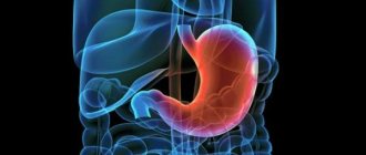
Not all beneficial substances entering the human body with food are absorbed directly in the stomach. Thus, water, sugar, and salt are partially absorbed. However, the main task of the stomach is precisely the digestion of food, or rather its processing into a pulp, the so-called chyme. How does this process work?
The digestion process begins even before food enters the stomach. As soon as a person thinks about food, sees it or smells it, the brain sends a signal to actively produce gastric juice. (Image 1).
The volume of an empty stomach of an adult is on average 0.5 liters. When food enters the lower esophagus, the inlet sphincter of the stomach, located in the cardia, opens and food enters. (Image 2). When filled with food, the stomach can stretch up to 1 liter, and in some people - up to 4 liters. It depends on the physiological characteristics of a person, his lifestyle and eating habits.
Then the muscle tissue of the stomach begins to contract, due to which the food is mixed with gastric juice and begins to be digested into chyme. Proteins and complex carbohydrates are broken down in the stomach (simple carbohydrates are broken down in the mouth under the influence of enzymes contained in saliva). Proteins, under the influence of enzymes, break down into amino acids, from which later the proteins that the body currently needs will be assembled. Complex carbohydrates break down into glucose, the simplest sugar. Later (but no longer in the stomach), glucose will break down into ATP (adenosine triphosphoric acid) molecules, the main source of energy for the human body. On average, food stays in the stomach for 3-4 hours. (Image 3).
When the work of the stomach is completed, the nervous system sends impulse signals to its muscle tissues, the lower sphincter - the pylorus - opens, chyme enters the duodenum, and the digestion process continues in the intestine. (Image 4).