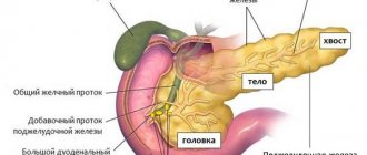
Treatment tactics for developed paraproctitis
Paraproctitis (especially acute) is an absolute indication for surgery.
Acute paraproctitis is a purulent inflammation of tissues. Any purulent focus in the body can resolve with several outcomes:
- The most favorable: the pus finds its way out on its own, the lesion is emptied, the wound heals, and self-healing occurs.
- The pus does not come out, but inside, spreads through the tissues, melting all surrounding tissues and organs, entering the blood and spreading throughout the body. The prognosis is unfavorable.
- The purulent focus is not completely emptied, part of it is encapsulated, creating a chronic focus with constant recurrence.
- The pus can come out completely, but the passage through which it came out does not heal; infection from the environment also constantly gets inside through it. The result is also a chronic inflammatory process.
So, the first most favorable outcome for untreated paraproctitis is possible only in 10-15% of cases. This information is for those who refuse surgery in the hope that “everything will work out.”
Therefore, when a diagnosis of acute paraproctitis is made, one cannot delay the operation.
Symptoms
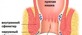
Fistulas are channels localized between the skin and the rectum, the external opening of which in most cases extends to the surface of the skin. In the absence of an external opening, an incomplete fistula with paraproctitis is diagnosed. Patients visiting a proctologist are interested in what a fistula looks like after paraproctitis. Most often with this disease, patients are concerned about the following manifestations:
- Fistula opening on the skin in the anus;
- Discharge of pus or blood from the opening;
- Itching and irritation caused by discharge;
- Painful sensations occur rarely, their cause is an exacerbation of the inflammatory process due to blockage of the fistula tract;
- Deterioration of the patient's general condition, decreased performance.
These manifestations of rectal fistula affect the daily life of patients, as they are forced to take measures to prevent the appearance of pus stains on clothes. In addition, their sleep is disturbed and regular headaches appear. If signs of paraproctitis appear, a consultation with a coloproctologist and subsequent diagnosis are necessary.
Long-term uncontrolled development of the disease can cause the following complications:
- regular exacerbations of the disease, in which the number of purulent cavities increases;
- scarring of soft tissues and the anal canal;
- changes in the sphincter muscles, resulting in decreased function;
- increasing the likelihood of malignant degeneration of affected tissues.
Timely treatment prevents negative consequences and helps improve the patient’s quality of life, therefore coloproctologists at the Yusupov Hospital provide medical care to patients with various forms of paraproctitis.
What does the pathological course look like?
Fistula with paraproctitis occurs as a result of a chronic inflammatory process in the tissue surrounding the rectum. The internal opening of the pathological tract is located in the rectum, through which the infection enters the tissue, which helps maintain the chronic inflammatory process.
What could be the consequences if paraproctitis is not operated on in time?
The consequences of refusing intervention and independent attempts to treat paraproctitis without surgery are as follows:
- Penetration of inflammation into deeper areas of tissue with the development of phlegmon.
- Pelvic pelvioperitonitis.
- Penetration of infection into the abdominal cavity with the development of peritonitis.
- Sepsis.
- Purulent melting of the pelvic organs - the walls of the rectum, bladder, urethra, genitals.
- Thrombosis and thrombophlebitis of the pelvic veins.
- Outcome into chronic paraproctitis.
Symptoms of paraproctitis
The onset of the inflammatory process can be marked by a slight increase in temperature (up to 37°C). When inflammation moves into the cellular space, i.e. In the development of acute paraproctitis itself, a complex of symptoms simultaneously appears. First of all, this is:
- temperature rise to 38°C (or higher);
- chills;
- pain in the rectal area not associated with bowel movements.
Subcutaneous paraproctitis has its own pronounced symptoms: the area of inflammation located next to the anus is distinguished by redness, swelling and thickening of the tissue. When palpated, a sharp pain is experienced.
With other types of paraproctitis, urination disorder, stool retention, and false urge to defecate may be observed.
Inflammation leads to the melting of cellular tissue and the accumulation of pus. In the absence of timely treatment, the resulting abscess breaks through and a fistula opening is formed - either outward (in the perineum, on the anterior abdominal wall or thigh), or into the intestinal lumen. In women, the abscess can open, forming a fistula in the vagina. The most dangerous option is the opening of a fistula into the abdominal cavity.
After the formation of a fistula, the bulk of the pus comes out, and the severity of the symptoms decreases. However, purulent processes continue, the disease becomes chronic.
Symptoms of chronic paraproctitis
With chronic paraproctitis, pain and discomfort are felt only if the exit of pus through the fistula is difficult for any reason. Usually pain is observed only with an internal fistula. The pain intensifies during bowel movements, and then subsides, since stretching of the intestines during the passage of stool promotes the release of pus. Pus or ichor is constantly secreted. With a rectal fistula, which has an external opening in the perineal area, the discharge irritates the skin, causing itching.
Chronic paraproctitis is characterized by a wave-like course of the disease. From time to time, the fistula canal becomes blocked by granulations and becomes clogged with dead tissue, as a result, pus again begins to accumulate in the area of inflammation and the symptoms of acute paraproctitis return. Then the abscess breaks out again and the severity of the symptoms disappears.
Stages of surgery for acute paraproctitis
- Opening and cleansing of the purulent focus.
- Elimination of the connection of the purulent focus with the rectum.
If both stages are completed, we can talk about radical surgery; complete recovery occurs in 80-85%.
However, it is not always possible to perform a radical operation at once. Opening a purulent focus during paraproctitis should be carried out as early as possible; this is an emergency situation, such intervention is carried out in the nearest surgical hospital.
Excision of the purulent tract and affected crypt requires the skill of a coloproctologist surgeon and should be carried out in a specialized proctology department. Often the second stage of the operation is carried out some time after the first.
General overview of the operation
Autopsy of acute paraproctitis is an emergency operation performed for life-saving reasons. Therefore, preparation for it is minimal, and there is only one contraindication - the patient’s extremely serious condition.
The opening of paraproctitis is usually carried out under general or epidural anesthesia, since it requires maximum muscle relaxation.
The easiest way to open ulcers is with superficial paraproctitis - subcutaneous and submucosal. They are also the most easily diagnosed - a general examination and rectoscopy (examination of the rectum using a rectal speculum) are sufficient.
Incisions used for acute paraproctitis: 1 – perianal abscess; 2 – retrorectal; 3 – ischiorectal
For subcutaneous paraproctitis, a semilunar incision is made around the anus in the place of greatest fluctuation and translucency of pus. The pus is released, all the bridges that divide the purulent cavity into several sections are destroyed. The purulent cavity is cleaned as much as possible, washed with antiseptics and antibiotics, and the wound is drained. Tamponade with antiseptic ointments (Levosin, Levomekol, Vishnevsky ointment) is possible.
With a highly qualified surgeon, the second stage can be carried out simultaneously: excision of the purulent tract that goes into the rectum. To do this, a button-shaped probe is inserted into the wound, with the help of which the passage is found. On the side of the rectum, at the site of the protruding end of the probe, the affected crypt is found. It is excised to healthy tissue. As a rule, sutures are not placed on the intestinal wall.
If the surgeon is unsure, the second stage of the operation can be delayed by 1-2 weeks (this is the period during which the purulent wound will be maximally cleaned and begin to heal, but it will still be possible to find its internal opening in the wall of the rectum. In addition, illiterate one-step excision of the external sphincter can lead to to its insufficiency in the postoperative period.
For submucosal paraproctitis, the incision is made from the side of the rectum. First, a digital examination and examination of the rectum in the speculum are performed. A puncture needle is inserted into the site of the largest protrusion. When pus is obtained, an incision is made in this place. Then, using a forceps, they pass bluntly into the cavity of the abscess; if necessary, the incision is widened. A rubber drainage is inserted into the opened abscess and its end is brought out through the anus.
The greatest difficulties are presented by surgery for ischiorectal, pelvic-rectal and retrorectal paraproctitis. The pus in these forms is localized deeply. Deep forms of paraproctitis are not always quickly diagnosed. To clarify the diagnosis and precise localization, a CT or MRI of the pelvic area is sometimes required.
The choice of access method for such paraproctitis is always difficult for the surgeon. Here, percutaneous access and emptying of the abscess with subsequent excision of the purulent tract or opening of the abscess only from the rectum is possible.
The fistula tract is inspected. If it is located transsphincterically, it is dissected into the rectal cavity using a probe, as with subcutaneous paraproctitis.
If the fistula tract is located extrasphincterically, it is usually excised with partial sphincterotomy (sphincter dissection) or the fistula tract is eliminated using the ligature method.
The essence of the ligature method is that a strong thread is inserted into the fistula tract. The incision is extended so that the thread is placed along the anterior or posterior midline of the sphincter. The thread is tied. Subsequently, every 2-3 days during ligation, the thread is tightened more and more tightly, which leads to a gradual crossing of the sphincter and elimination of the fistula tract. Such a gradual, rather than sudden, dissection of the sphincter allows one to avoid the formation of its insufficiency after surgery.
Description of the stages of the operation
- The stage of opening and releasing pus from the abscess in the rectum is performed using epidural or local anesthesia, since surgical intervention requires the most relaxed anal sphincter. For subcutaneous paraproctitis, the surgeon makes an external semicircular incision around the anus in the place where the cavity is palpated or visualized. The doctor dissects all the bridges that divide the crypt cavity, cleans and rinses it with a disinfectant solution and drains it to release the exudate.
- The second stage is the detection and resection of the duct that connects the pouch and the rectal cavity. Using a probe inserted into the crypt cavity, a duct and its exit into the intestine are detected. Then the passage is excised. No stitches are required. When the abscess is localized in the submucosal layer, access to it is made from the rectal cavity. The doctor inserts a rectal speculum into the intestine and finds a protrusion that forms a purulent cavity. A needle is inserted into it, and if pus appears during puncture, the abscess is opened, cleaned, and drainage is removed through the anus.
The most difficult type of surgical intervention is surgery for pus in the crypt, localized in the following area:
- ischiorectal;
- retrorectal;
- pelvic-rectal.
Precise localization of the abscess is difficult, since the crypts are located in deep layers of tissue. They are detected during instrumental examination - using CT or MRI. The abscess is then opened using a percutaneous or intraintestinal approach. If the abscess is localized in the space outside the sphincter, then the approach to the cavity and resection of the canal is performed percutaneously.
If the inflamed crypt is located deep in the tissue behind the anal sphincter, then access is made from inside the rectum. In this case, partial dissection of the sphincter ring can be performed. The move is removed using a ligature.
With this method, a ligature thread is inserted into the fistula tract, the ends are brought out from the sphincter and from its outer side, and the thread is tied into a ring. The thread is tightened daily, gradually cutting the sphincter.
This method avoids the risk of relapse of the pathological process as a result of insufficiency of the fistula tract and incompetence of the anal sphincter. It is believed that in the intervals between tightening the ligature, the wound gradually scars and when the thread completely cuts through the anal sphincter, a thin scar is formed that does not affect the obturator function.
Chronic paraproctitis
Chronic paraproctitis occurs after spontaneously opened or inadequately treated paraproctitis. In 10-15% of cases, it can occur after proper adequate drainage of acute paraproctitis.
Actually, chronic paraproctitis is a fistula that occurs in the soft tissues of the peri-rectal region. It can be complete (with two outlet openings - in the skin of the perineum and in the wall of the rectum) and incomplete (one opening is external or internal). It can also have multiple branches and multiple holes.
The presence of a fistula implies constant infection from the environment and constant recurrence of inflammation in the peri-rectal tissue.
Treatment of chronic paraproctitis is surgical. The operation can be either emergency (during exacerbation of the disease) or planned.
The most favorable prognosis is to perform a planned operation in subacute cases after some preparation (anti-inflammatory and antibacterial therapy). It is not recommended to perform the operation during a period of stable remission, since the internal opening of the fistula may not be found at this time.
Treatment of paraproctitis
Acute paraproctitis
If acute paraproctitis is diagnosed, surgery is performed urgently after short preoperative preparation. During surgery for acute paraproctitis, the surgeon, under general anesthesia, performs a wide opening and drainage of the abscess with removal of damaged tissue. It is highly desirable to open the abscess with simultaneous dissection of the purulent tract and excision of the inflamed crypt, which is the entry point for the infection. Otherwise, if we limit ourselves to only opening the abscess, then the patient will either form a fistula in the near future, or there will be a relapse (recurrence) of acute paraproctitis. It should be noted that performing extended operations (with excision of the crypt) is difficult in conditions of purulent inflammation, however, as a rule, a proctologist with proper experience can do this. The operation ends with drainage. After the operation, the patient is prescribed individual therapy associated with the administration of antibacterial drugs, infusion, detoxification therapy, the administration of analgesics and daily changes of ointment dressings.
Chronic paraproctitis
Treatment of chronic paraproctitis is aimed at excision of the fistula tract with mandatory removal of the internal opening of the fistula. In some cases, a mucous flap of the rectum is moved to the site of the eliminated internal opening, which prevents the penetration of infection into the peri-intestinal tissue (and, accordingly, prevents microbes from entering the perirectal tissue) (Fig. 5). Sometimes, if the fistula is located in the muscle fibers of the sphincter, it is necessary to disconnect the rectal muscles and then restore them. Given the important physiological role of the sphincter, the operation should be performed by a proctologist with experience in such interventions. In our clinic, both electrosurgical instruments and modern Liga Sure ultrasonic knives are used to perform such operations. These instruments not only make it easier for the surgeon to perform the operation, but also reduce tissue trauma, which, in turn, reduces the risk of damage to the rectal muscles, reduces pain and reduces the recovery period after surgery.
In the last decade, laser technology (LHP) has been actively introduced in the surgical treatment of rectal fistulas. The principle of the operation is the thermal effect of a laser beam on the wall of the rectal fistula, during which its coagulation and sterilization occurs (Fig. 6).
Such interventions are characterized by less intense pain in the postoperative period and a short period of hospitalization. The advantages of the technique are:
- lack of a wide incision;
- reducing the risk of suppuration;
- minimizing trauma to the rectal sphincter.
However, good “stable” results with this treatment can be obtained, as a rule, with “low” fistulas located medially from the sphincter.
In any case, in our Clinic the choice of method of surgical intervention is made by a surgeon - a coloproctologist with experience in treating such diseases, with maximum consideration for the interests of the patient.
Types of operations for chronic paraproctitis
The main goal of surgical intervention for chronic paraproctitis is the elimination of the fistula tract. The extent of the operation depends on the location of the fistula.
To accurately localize the openings of the fistula tracts, dyes (methylene blue) are injected into the wound. Sometimes radiopaque contrast is used with radiography.
Types of operations for chronic paraproctitis:
- Dissection of the fistula.
- Fistula excision.
- Ligature method.
- Plastic surgery.
- Laser obliteration of fistula.
- Obliteration of the fistula with a collagen thread.
With a transsphincteric location of the fistula, it is possible to dissect the fistula tract from the rectal lumen or excise it (Gabriel's operation) along its entire length, followed by complete or partial suturing of the wound.
In case of extrasphincteric location of the fistula (after pelvic-rectal or ischiorectal paraproctitis), excision of the fistula is performed with dosed sphincterotomy or the ligature method.
Plastic surgery involves excision of the fistula and closing its internal opening with a flap of the intestinal mucosa.
New methods - laser coagulation of the fistula tract or filling it with collagen thread - are possible if the fistula tract has a simple linear shape.
LIFT operation with laser assistance for rectal fistulas
The peculiarity of this operation is that the fistula tract is processed and closed from access in the intersphincteric space. Those. the surgeon does not cross the sphincters, but goes between them to close the fistulous tract. The outer part of the fistula tract is treated with laser coagulation, which allows the removal of necrotic and infected tissue.
Click to view photos. Attention! Profile surgical photo, 18+.
Rice. 3. Fistula of the rectum. LIFT operation with laser assistance
After operation
After surgery for acute or chronic paraproctitis, it is important to follow some rules. It is advisable to spend the first few days, even after opening the superficial paraproctitis, in a hospital. Antibiotics and painkillers are prescribed. Dressings are performed daily and can be quite painful.
Immediately after the operation, a slag-free diet is prescribed - semolina or rice porridge with water, steamed meatballs, boiled fish, steamed omelettes. It is necessary to retain stool for 2-3 days after surgery.
After 2-3 days, if there is no independent stool, a cleansing enema is given. It is very important to prevent both constipation and diarrhea. Normal stool has no effect on wound healing. Gradually, baked apples, boiled vegetables, a decoction of dried fruits, and lactic acid products are added to the diet. It is important to drink at least 5 glasses of liquid per day.
Spicy, salty foods and alcohol are absolutely excluded. You should refrain from raw vegetables and fruits, legumes, baked goods, whole milk, and carbonated drinks.
If the postoperative period is normal, the patient can be sent home after a few days. He can carry out further dressings himself. They usually involve treating the wound with hydrogen peroxide, then washing it with an antiseptic (chlorhexidine, miramistin or furatsillin solution) and applying a sterile napkin with antibacterial ointment.
After each stool, a thorough toilet of the perineum is necessary, sitz baths and a new dressing are desirable. In case of stool retention, microenemas can be used.
At first, purulent contents, ichor, will flow out of the wound. Sanitary pads will be required. Over time, the discharge from the wound will become less and less.
The period of incapacity for work after an uncomplicated operation is about 8-10 days. Complete healing of a purulent wound usually occurs after 3-4 weeks.
The patient is also warned that partial insufficiency of the anal sphincter may persist for 1-2 months after surgery. This may manifest itself in periodic incontinence of gas and loose stools. For prevention, special gymnastics for the sphincter are prescribed.
Paraproctitis surgery
Since the disease is very severe and there is no opportunity for conservative treatment, the only option is surgery. After diagnosis, it is necessary to carry out intervention as soon as possible if paraproctitis is acute. However, it is also important to treat paraproctitis after surgery and follow all instructions.
If there is no relapse, then conservative treatment is possible. Typically, non-hot sitz baths with medicinal solutions, compresses, microenemas and antibiotics are prescribed.
Preoperative preparation
To determine the progress of the procedure, it is necessary to find out the nature of the course of acute paraproctitis. A favorable course, according to statistics, occurs only in 10-15% of cases. In total, there are 4 main currents among them:
- the pus comes out and self-healing occurs;
- pus comes out and inside, surrounding tissues and organs become inflamed, blood poisoning occurs;
- the focus with pus is not completely emptied and constant chronic inflammation occurs;
- the pus comes out completely, but the tract and the lesion themselves become inflamed.
Preparation for paraproctitis surgery does not take much time and does not require serious manipulation. Children under one year of age are prescribed a cleansing enema no more than a day before surgery. In this case, there must be no exacerbation of paraproctitis.
For older children and adults, warm sitz baths are prescribed as preoperative preparation 2 times a day for 3-5 days before surgery. If a fistula has formed, its course is washed with a solution of furatsilin or rivanol.
On the day of the operation, cleansing enemas are given, and the evening before with an antiseptic solution. The diet should contain fermented milk products. You need to exclude meat, legumes, and some vegetables. It is necessary to follow a diet after paraproctitis surgery.
If the course of the disease is acute, then patients need to undergo a course of antibacterial and anti-inflammatory therapy. After the inflammation has subsided, surgery should be performed as soon as possible to avoid relapse.
In cases such as old age, weakened immunity, severe illness, surgery is not always possible. Therefore, they try to improve the condition with conservative treatment, and only then operate.
There are times when conservative methods provide good enough results to postpone surgery.
Progress of the operation
After all the preparatory procedures have been completed, an operation is prescribed, which lasts no more than 30 minutes. Anesthesia is used only sacral or epidural. The operation to treat paraproctitis involves two steps: excision of the abscess or opening and drainage of the abscess.
To understand how long paraproctitis takes to heal after surgery, it is necessary to take into account the age and individual characteristics of the body.
An urgent operation to open acute paraproctitis is performed only if there is evidence for it. The procedure cannot be performed if the tissue around the surgical incision site is inflamed and if the location of the anal sinus is unknown.
Surgery for acute paraproctitis can be done in three ways:
- First, the abscess is opened , drained, then the sinuses and the purulent tract into the rectum are excised. This option is appropriate if the passage is located inside the external sphincter and if only the subcutaneous layer is affected.
- In case of intersphincteric inflammatory process, the abscess is opened, the contents are drained , the anal sinus is excised and a sphincterotomy is performed.
- For trans- and extrasphincteric paraproctitis, the abscess is cut, the purulent masses are removed, the crypts are excised, and a ligature is applied for drainage. As a result of this operation, there is a good outflow of content.
Most often, a multi-stage operation is performed, which involves several stages. At first, the abscess is opened and the contents are removed. At the next stage, after about 5-7 days (depending on tissue healing), the anal sinus and glands are removed. The technique for opening subcutaneous paraproctitis depends on the nature of the inflammation.
After operation
Treatment of acute paraproctitis after surgery includes complex therapy. If there are no complications and healing proceeds normally, then the patient is sent home after a few days.
Healing itself after an uncomplicated operation lasts up to 10 days, and a purulent wound heals completely only within 4 weeks.
Antiviral and antibiotics are almost always prescribed to avoid the inflammatory process. Cleansing enemas with medications are also required. Ointments must be constantly applied to the wound to heal.
If stool retention occurs after 2-3 days, then cleansing enemas are used. After each trip to the toilet, you need to treat the wound : sitz baths and a new dressing.
Don't hesitate to see a doctor
Often, when pain occurs in the anus, patients do not rush to see a doctor because they are embarrassed to show the doctor their private parts. They self-medicate, buying ointments and suppositories for hemorrhoids in pharmacies, and use dubious recipes from the Internet. All this only aggravates the situation and can lead to complications.
In addition, all this time I have to endure really very strong and growing pain. According to reviews from patients who have undergone surgery, after opening the abscess, the severe pain goes away almost immediately.
Summarizing all of the above, we must say to those who doubt and feel embarrassed: if pain in the anus appears in combination with fever and general malaise, you should consult a doctor as soon as possible, preferably a coloproctologist.
Paraproctitis is a formidable disease, difficult to treat even in the initial stages. The consequences may be irreversible.
Cost of surgery for paraproctitis
Opening and draining an abscess of peri-intestinal tissue can be performed urgently and free of charge in any surgical department. Of course, it is advisable, even in an emergency situation, to go to a specialized department, where they can simultaneously perform a radical operation - that is, the elimination of a purulent tract.
If it is impossible to do this, you will have to undergo a repeat operation of excision of the crypt in the coloproctology department.
Prices in paid clinics:
- Opening an abscess - from 5,000 rubles.
- Radical surgery for acute paraproctitis – from 16,000 rubles.
- Excision of rectal fistula – from 12,000 rubles.
- Excision of chronic paraproctitis with a laser – from 15,000 rubles.
Laser coagulation of rectal fistula
Laser coagulation of a rectal fistula allows you to remove the fistula without damaging the anal sphincters. During the operation, the laser conductor is inserted into the fistula tract along its entire length from the external fistula opening to the internal one. Laser energy is then supplied to the light guide. Radial dispersion of the laser beam allows uniform coagulation of all walls of the fistula tract, leading to photothermal destruction of the fistula and collapse. As a result of the manipulation, the lumen of the fistula tract collapses.