Classifications of GERD
Much in therapy depends on the stages of the disease. Such information influences the duration of treatment and the choice of certain drugs. In the case of GERD, what matters first is how deeply the esophageal mucosa is affected. In medicine, the classification of gastroesophageal reflux disease is more often used, which is detected by a research method such as FGDS (fibrogastroduodenoscopy).
What symptoms will bother a person at each stage of the disease? Today we have to answer not only this question. There are several options for classifying GERD; let’s look at the most common ones.
Classification of pathology by degree of development
If the pathology is not treated, it will progress. In its development it has several stages. The classification of GERD in this case is as follows:
- first degree - the last areas are characterized by redness of the tissues, slight erosions, although sometimes such signs cannot be detected);
- second stage – damage extends to more than 20% of the esophagus, the patient develops persistent heartburn;
- third degree - not only the upper layer of the mucous membrane is destroyed, but also deeper tissues; Ulcers appear that affect the muscles. The stage is characterized by burning, pain in the chest area, worse at night;
- fourth - characterized by damage to almost the entire surface of the mucous membrane, while the symptoms increase significantly;
- The fifth stage is the most severe form of pathology, in which various complications of GERD already appear.
Note! This classification is the most common and easiest to understand. On its basis, therapeutic measures are prescribed to help eliminate damage to the mucous membrane and symptoms.
Endoscopic classification of GERD
Endoscopic classification was proposed in the late 80s by Savary and Miller, and is quite widely used in our time.
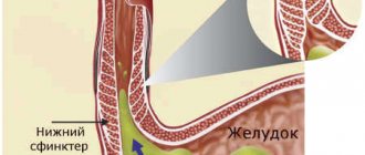
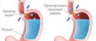
It has long been known that the mechanism of development of GERD is a dysfunction of the lower esophageal sphincter (a muscle located on the border between the esophagus and the stomach, limiting the reverse movement of food). When this muscle is weakened, gastric contents, including hydrochloric acid, are refluxed into the esophagus. And over time, almost all of its shells undergo changes. So they served as the basis for this classification.
How the disease develops
Statistics show that almost half of the adult population has manifestations of gastro-food reflux. Of this number, 10% of people showed endoscopic signs of the disease. This suggests that the mechanism of development of this disease passes quite unnoticed.
heartburn or nausea after eating , but they see no point in going to see a doctor. Often this disease of the esophagus is diagnosed as a result of the development of more complex inflammatory processes in the gastrointestinal tract.
Important! The main mechanism that triggers the development of gastro-food reflux is impaired motility.
The stomach begins to throw its contents into the esophagus, and particles of the contents are excreted from there for a long time. Until the esophagus copes with this reflux, the person will feel pain. Nature has given our body several protective functions against the occurrence of reflux.
First, the lower esophageal sphincter must establish an anti-reflux barrier in a timely manner.
If relaxation of this part of the esophagus occurs for a long time, then its mucous membrane is longer exposed to the negative effects of acids.
Secondly, saliva is able to neutralize the negative effects of hydrochloric acid, which is important when throwing stomach contents into the esophagus. In people who already have developed reflux esophagitis, doctors note unsatisfactory gastric motility and disruptions in the volume of salivation.
Los Angeles classification of GERD
At the end of the 20th century, at the European Gastroenterology Week, it was proposed to use the extent of lesions. This is how the Los Angeles classification of GERD was born. Here's what it includes.
- Severity grade A - there is one or more lesions of the esophageal mucosa (erosions or ulcers), each of which is no more than 5 mm, within only one fold of the mucous membrane.
- Grade B - the changes also affect only one fold, but one of the lesions may extend beyond 5 mm.
- Grade C - the process has already spread to 2 folds or more, areas with changes of more than 5 mm. At this stage, damage to the esophagus reaches 75%.
- Grade D - most of the esophagus is affected. The lesion circumference is at least 75%.
According to the Los Angeles classification, complications in the form of ulcers and contractions can be present at any of the above stages.
Disease-stage units were created to simplify the work of doctors. Thanks to classifications, it becomes easier to understand the manifestations of the process and better select methods for its treatment. Only a doctor can determine at what stage of development of the disease each person suffering from GERD is. Therefore, at the first signs of illness, to speed up recovery, contact a specialist.
Clinics for treatment with the best prices
Price
Total: 475in 25 cities
Source
a common part
Gastroesophageal reflux disease (GERD)
is a disease characterized by the development of specific symptoms and/or inflammatory lesions of the distal esophagus due to repeated, retrograde flow of gastric and/or duodenal contents into the esophagus.
The pathogenesis is based on insufficiency of the lower esophageal sphincter (circular smooth muscle, which is in a state of tonic contraction in a healthy person and separates the esophagus and stomach), which contributes to the reflux of stomach contents into the esophagus (reflux).
Long-term reflux leads to esophagitis and sometimes to tumors of the esophagus. Typical (heartburn, belching, dysphagia) and atypical (cough, chest pain, wheezing) manifestations of the disease occur.
Pathological changes in the respiratory system (pneumonia, bronchospasm, idiopathic pulmonary fibrosis), vocal cords (hoarseness, laryngitis, laryngeal cancer), hearing organ (otitis media), teeth (enamel defects), may be additional signs indicating reflux .
The diagnosis is made based on a clinical assessment of the symptoms of the disease, the results of endoscopic studies, and pH-metry data (monitoring pH in the esophagus).
Treatment consists of lifestyle changes and taking medications that reduce gastric acidity (proton pump inhibitors). In some cases, surgical treatment methods may be used.
- Classification of GERD
First of all, the classification divides gastroesophageal reflux disease into 2 categories: GERD with esophagitis and GERD without esophagitis.
GERD with esophagitis (endoscopically positive reflux disease)
Reflux esophagitis is damage to the mucous membrane of the esophagus, visible during endoscopy, an inflammatory process in the distal (lower) part of the esophagus caused by the action of gastric juice, bile, pancreatic and intestinal secretions on the mucous membrane of the esophagus. It is observed in 30-45% of patients with GERD.Complications of reflux esophagitis are:
- Esophageal strictures.
- Barrett's esophagus.
- Adenocarcinoma of the esophagus.
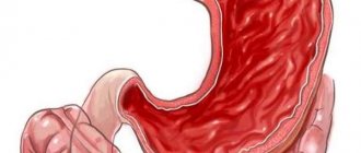
Erosion and ulcers of the esophagus, accompanied by bleeding.
The condition of the esophageal mucosa is assessed endoscopically according to the M.Savary-J.Miller classification, or according to the Los Angeles (1994) classification.
- Classification by M.Savary-J.Miller as modified by Carrison et al. Grade 0 – there are no signs of reflux esophagitis.
- I degree – non-confluent erosions against the background of mucosal hyperemia, occupying less than 10% of the circumference of the distal esophagus.
- Grade II - confluent erosive lesions occupying 10-50% of the circumference of the distal esophagus.
- III degree – multiple, circular erosive and ulcerative lesions of the esophagus, occupying the entire circumference of the distal esophagus.
- IV degree – complications: deep ulcers, strictures, Barrett’s esophagus.
- Grade A - one or more defects of the esophageal mucosa no more than 5 mm long, none of which extends to more than 2 folds of the mucous membrane.
- GERD without esophagitis (endoscopically negative reflux disease, or non-erosive reflux disease)
GERD without esophagitis (endoscopically negative reflux disease, or non-erosive reflux disease) is damage to the esophageal mucosa that is not detected by endoscopic examination. Occurs in more than 50% of cases.The severity of subjective symptoms and duration of the disease do not correlate with the endoscopic picture. With endoscopically negative GERD, quality of life suffers in the same way as with reflux esophagitis, and pH measurements characteristic of the disease are observed.
- Epidemiology of GERD
The incidence of GERD is often underestimated, since only 25% of patients consult a doctor. Many people do not complain because they manage the symptoms of the disease with over-the-counter medications. The occurrence of the disease is promoted by a diet containing excess amounts of fat.If we evaluate the prevalence of GERD by the frequency of heartburn, then 21-40% of residents of Western Europe, up to 20-45% of residents of the USA and about 15% of residents of Russia complain about it. The likelihood of having GERD is high if you experience heartburn at least twice a week. In 7-10% of patients it occurs daily. However, even with more rare heartburn, the presence of GERD cannot be excluded.
The incidence of GERD in men and women of any age is (2-3):1. Incidence rates of GERD increase in people over 40 years of age. However, Barrett's esophagitis and adenocarcinoma are approximately 10 times more common in men.
- ICD 10 code K21.
Source
Before you find out how GERD is classified according to ICD 10 code, you need to consider what kind of disease it is.
It is a lesion of the mucous membrane of the esophagus. The abbreviation can be deciphered as follows: gastroesophageal reflux disease.
It is characterized by periodic reflux of stomach contents back into the esophagus. In this case, the sphincter is affected and inflammation develops.
What is GERD and the disease code according to ICD-10?
Treatment of GERD includes lifestyle changes, drug therapy, and in the most difficult cases, surgery. Drug therapy for GERD and lifestyle changes for patients with GERD are aimed at treating inflammation of the esophageal mucosa, reducing the number of gastroesophageal refluxes, reducing the damaging properties of refluxate, improving the cleansing of the esophagus from aggressive gastric contents that have entered it and protecting the esophageal mucosa. [2]
Lifestyle change
Patients suffering from GERD are recommended to:
- Normalization of body weight.
- Eliminate smoking, reduce consumption of alcohol, fatty foods, coffee, chocolate, carbonated drinks.
- Eating small portions regularly, up to five times a day; dinner no later than 2-3 hours before bedtime.
- Avoiding stress associated with increased intra-abdominal pressure, as well as wearing tight belts, belts, etc.
- Elevated position (15-20 cm) of the head end of the bed at night.
Drug therapy
Drug therapy for GERD is mainly aimed at normalizing acidity and improving motility. Antisecretory agents (proton pump inhibitors, H2-histamine receptor blockers), prokinetics and antacids are used to treat GERD.
Proton pump inhibitors (PPIs) are more effective than H2 blockers and have fewer side effects. It is recommended to take the PPI rabeprazole at a dose of 20-40 mg/day, omeprazole at a dose of 20-60 mg/day or esomeprazole at a dose of 20-40 mg/day for 6-8 weeks. [2] When treating erosive forms of GERD, PPIs are taken for a long time, several months or even years. In this situation, the issue of IPN security becomes important. Currently, there are suggestions of an increase in bone fragility, intestinal infections, community-acquired pneumonia, and osteoporosis. During long-term treatment of GERD with proton pump inhibitors, especially in elderly patients, interactions with other medications often have to be taken into account. If it is necessary to take other drugs simultaneously with PPIs for the treatment or prevention of other diseases, preference is given to pantoprazole, as it is the safest in terms of interactions with other drugs. [3]
Treatment
Non-drug therapeutic measures for gastroesophageal disease: • normalization of body weight, adherence to a diet (small portions every 3-4 hours, eating no later than 3 hours before bedtime), avoidance of foods that help relax the esophageal sphincter (fatty foods, chocolate , spices, coffee, oranges, tomato juice, onions, mint, alcohol-containing drinks), increasing the amount of animal protein in the diet, avoiding hot food and alcohol; • it is necessary to avoid tight clothing that pinches the torso; • it is recommended to sleep on a bed with the head of the bed raised by 15 centimeters; • to give up smoking; • it is necessary to avoid prolonged work in an inclined position, heavy physical exertion; • medications that negatively affect esophageal motility (nitrates, anticholinergics, beta-blockers, progesterone, antidepressants, calcium channel blockers), as well as non-steroidal anti-inflammatory drugs that are toxic to the esophageal mucosa are contraindicated. Drug treatment of gastroesophageal reflux disease is carried out by a gastroenterologist. Therapy takes from 5 to 8 weeks (sometimes the course of treatment reaches a duration of up to 26 weeks), is carried out using the following groups of drugs: antacids (aluminum phosphate, aluminum hydroxide, magnesium carbonate, magnesium oxide), H2-histamine blockers (ranitidine, famotidine), proton pump inhibitors (omeprazole, rebeprazole, esomeprazole). In cases where conservative therapy for GERD does not produce an effect (about 5-10% of cases), or when complications or a diaphragmatic hernia develop, surgical treatment is performed. Surgical interventions for gastroesophageal reflux disease: endoscopic plication of the gastroesophageal junction (sutures are placed on the cardia), radiofrequency ablation of the esophagus (damage to the muscular layer of the cardia and gastroesophageal junction, in order to scar and reduce reflux), gastrocardiopexy and laparoscopic Nissen fundoplication.
K20-K31 Diseases of the esophagus, stomach and duodenum
Excluded:
diaphragmatic hernia (K44.-)
(+) The following subcategories are intended for use with categories K25-K28:
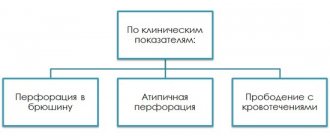
.0 - acute with bleeding .1 - acute with perforation .2 - acute with bleeding and perforation .3 - acute without bleeding or perforation .4 - chronic or unspecified with bleeding .5 - chronic or unspecified with perforation .6 - chronic or unspecified with bleeding and perforation .7 — chronic without bleeding or perforation .9 — not specified as acute or chronic without bleeding or perforation
Included:
Esophageal abscess Esophagitis:
If necessary, identify the cause, use an additional external cause code (class XX)
Excluded
: esophageal erosion (K22.1), reflux esophagitis (K21.0) esophagitis with gastroesophageal reflux disease (K21.0)
K21 Gastroesophageal reflux disease
K21.0 Gastroesophageal reflux disease with esophagitis
K22 Other diseases of the esophagus
Excluded:
varicose veins of the esophagus (I85.-)
K22.0 Achalasia cardia
Excluded:
congenital cardiospasm (Q39.5)
K22.1 Esophageal ulcer
Erosion of the esophagus Esophageal ulcer:
- NOS
- caused by: chemicals
- medicines and medicines
- fungal
- peptic
If necessary, identify the cause, use an additional external cause code (class XX)
K22.2 Esophageal obstruction
Esophageal compression Esophageal narrowing Esophageal stenosis Esophageal stricture
Excluded:
congenital stenosis and stricture of the esophagus (Q39.3)
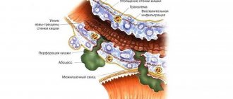
K22.3 Perforation of the esophagus
Excluded
: traumatic perforation of the (thoracic part) of the esophagus (S27.8)
K22.4 Esophageal dyskinesia
K22.5 Esophageal diverticulum
Excluded
: congenital esophageal diverticulum (Q39.6)
K22.6 Gastroesophageal rupture hemorrhagic syndrome
Excluded
: Barrett's ulcer (K22.1)
K22.8 Other specified diseases of the esophagus
K22.9 Disease of the esophagus, unspecified
K23.0* Tuberculous esophagitis (A18.8 +)
K23.8* Lesions of the esophagus in other diseases classified elsewhere
K25 Stomach ulcer
See (+) subcategories at the beginning of the page
erosion (acute) of the stomach, peptic ulcer:
- pyloric part
- stomach
If it is necessary to identify the drug that caused the injury, use an additional code for external causes (class XX)
acute hemorrhagic erosive gastritis (K29.0) peptic ulcer ulcer NOS (K27)
erosion (acute) of the duodenum, peptic ulcer:
- duodenum
- postpyloric part
If it is necessary to identify the drug that caused the injury, use an additional code for external causes (class XX)
Excluded:
peptic ulcer NOS (K27)
K27 Peptic ulcer of unspecified localization
See (+) subcategories at the beginning of the page
Included:
gastroduodenal ulcer NOS peptic ulcer NOS
Excluded:
peptic ulcer of the newborn (P78.8)
K28 Gastrojejunal ulcer
See (+) subcategories at the top of the page
Included:
peptic ulcer or erosion:
- anastomosis
- gastrocolic
- gastrointestinal, gastrojejunal
- jejunal
- regional
- anastomosis
Excluded:
primary ulcer of the small intestine (K63.3)
eosinophilic gastritis or gastroenteritis (K52.8) Zollinger-Ellison syndrome (E16.8)
Acute (erosive) gastritis with bleeding
Excluded:
erosion (acute) of the stomach (K25.-)
K29.2 Alcoholic gastritis
Atrophy of the gastric mucosa
Hypertrophic giant gastritis Granulomatous gastritis Menetrier's disease
K29.7 Gastritis, unspecified
K29.9 Gastroduodenitis, unspecified
Included:
digestive disorders
K31 Other diseases of the stomach and duodenum
Included:
functional stomach disorders
duodenal diverticulum (K57.0-K57.1) gastrointestinal bleeding (K92.0-K92.2)
Excluded:
congenital or infantile pyloric stenosis (Q40.0)
K31.2 Hourglass stricture and gastric stenosis
congenital hourglass-shaped stomach (Q40.2) hourglass-shaped gastric constriction (K31.8)
K31.3 Pylorospasm, not elsewhere classified
How does a doctor diagnose reflux esophagitis?
Any diagnosis of the disease should begin with a consultation with a doctor. The doctor will clarify the nature of the pain, its frequency and duration. The doctor can also find out the patient’s dietary habits to determine his lifestyle. After the conversation, the doctor may examine the tongue.
With gastrointestinal reflux, the tongue may be coated with a white coating. To rule out other diseases, the doctor should palpate the abdomen.
If no painful sensations are detected, then the patient is referred for an instrumental examination.
Using a probe and a camera at its end, you can see a clear picture of the gastrointestinal tract disease. With reflux, the lining of the esophagus will be red. In some cases, your doctor may order tissue to be removed from the area for further testing.
Also used for diagnosis:
- x-ray,
- daily pH-metry (determination of acidity level),
- esophagomanometry (determining the functionality of the lower esophageal sphincter),
- ECG (to rule out heart disease),
- Chest X-ray (to rule out lung diseases).
Taken together, all diagnostics will allow you to see an accurate picture of the course of the disease. The main thing is to see a doctor in time.
Definition of disease and code according to ICD-10
What is duodenogastric reflux (DGR): this term refers to the pathological reflux of bile, gastric and pancreatic juices into the stomach and lumen of the esophagus due to weakness of the obturator sphincters.
Normally, digested and crushed food (chyme) enters the lumen of the small intestine through the pyloric section of the stomach, which is represented by a powerful circular muscle - the pyloric sphincter. Its contraction prevents the backflow of intestinal contents.
Many scientists are inclined to believe that biliary reflux is not an independent disease, but a syndrome that occurs against the background of existing pathologies of the gastrointestinal tract. In some cases, it is classified as gastroesophageal reflux disease, in which the reflux of bile into the stomach is quite common.
The relevance of the problem lies not only in its high prevalence, but also in the fact that the presence of duodenogastric bile reflux contributes to the development of concomitant pathologies and a deterioration in the quality of life. If diagnosed untimely, GHD becomes chronic with frequent relapses, which ultimately leads to an increase in the duration and cost of treatment.
ICD-10 code
According to the tenth revision of the Board of the International Classification of Diseases, biliary reflux does not have its own ICD code, which once again confirms the secondary nature of its occurrence. The syndrome may be part of the following diagnoses:
- Gastroesophageal reflux disease (K.21).
- Duodenitis (K.29).
- Gastritis (K.29.3).
- Gastroduodenitis of unknown etiology (K.29.9).
Causes
Factors contributing to the development of GERD are: impaired motor functions of the upper digestive tract, hyperacidotic conditions, reduced protective function of the esophageal mucosa. Most often, with GERD, there is a violation of two nature-provided mechanisms for protecting the esophagus from the aggressive environment of the stomach: esophageal clearance (the ability of the esophagus to evacuate contents into the stomach) and the resistance of the mucous wall of the esophagus. Stress, smoking, obesity, frequent pregnancies, diaphragmatic hernia, medications (beta-blockers, calcium channel blockers, anticholinergics, nitrates) increase the likelihood of developing gastroesophageal reflux disease.
Causes of the disease and risk factors
The independent course of gastroduodenal reflux occurs in 25% of cases of all disorders of the valvular apparatus of the digestive organs. Otherwise, the pathology is due to the presence of other diseases of the gastrointestinal tract in the patient.
- chronic gastroduodenitis, gastritis;
- chronic pancreatitis and cholecystitis;
- functional dyspepsia and irritable bowel syndrome;
- peptic ulcer of the duodenum, stomach;
- giardiasis, helminthic infestation;
- congenital anomalies of the gastroduodenal zone.
The main causes of bile (alkaline) reflux also include:
- functional insufficiency and immaturity of the sphincter of the excretory section of the stomach (due to dysfunction of the nervous regulation of this area against the background of neuroses);
- deficiency of the stimulating hormone – gastrin;
- lack of consistency between the cycles of relaxation and contraction of the muscles of the stomach and small intestine;
- duodenal hypertension - high pressure in the lumen of the duodenum due to a hernia, curvature of the spinal column, tumor process or prolapse of internal organs;
- violation of peristalsis of the digestive system.
Among the provoking factors, it is worth noting old age, irregular rough eating, overeating, alcohol abuse, smoking, long-term and uncontrolled use of NSAIDs (non-steroidal anti-inflammatory drugs). An important role in the genesis of reflux is played by high acidity of gastric juice and previous operations on the stomach and intestines.
Features of classification by ICD code
Reflux esophagitis is a complex disease characterized by unpleasant symptoms and painful sensations. A person cannot eat what he wants, as this causes severe discomfort.
The pathology is manifested by heartburn, regurgitation, and bad breath. In some cases, there is an increase in temperature, the urge to vomit, and the inability to swallow food.
Classification of esophagitis will help determine the direction of treatment. The international disease code is K21.
However, this pathology can have various forms, which also need to be considered:
- ICD K-21. This is refractory GERD, in which the patient not only develops an inflammatory process in the sphincter area. Erosion appears on this part of the organ.
- K-21.2. In this case, the esophageal component is absent. That is, there are unpleasant symptoms, but they are not associated with damage to the inner surface of the esophagus, since there are none.
Clinical manifestations of the disease are present in both cases, but they are different. In the second case, there is no threat to life.
Important! The cause of GERD can be either a physiological or psychosomatic factor. The cause of the pathology must be determined before treatment is carried out.
The mechanism of development of duodenogastric reflux
The stomach, in turn, produces a hormone - gastrin, which regulates the peristalsis of the organ and has a direct effect on the tone of the pyloric sphincter.
Glucagon, cholecystokinin, secretin, histamine normalizes gastric motility. The work of the muscular apparatus of the digestive tube depends on their concentration. Hormonal imbalance, impaired nervous regulation - all this contributes to the occurrence of pathological reflux of duodenal contents into the stomach cavity, and often into the lumen of the esophagus.
Pregnancy is another common factor in the pathology. An enlarged uterus leads to an increase in intra-abdominal pressure in the abdominal cavity and compression of the duodenum, promoting regurgitation of bile and digestive enzymes upward and the appearance of symptoms.
Causes of reflux esophagitis in adults
To protect yourself from the occurrence of reflux esophagitis, you need to know the main risk factors for the development of this disease and the possible causes of its development. Experts note that the main factors that provoke the appearance of such an inflammatory process are:
- obesity;
- frequent vomiting;
- installation of a nasogastric tube (for enteral nutrition);
- pregnancy;
- hiatal hernia.
All this can provoke the appearance of reflux esophagitis. There are a number of reasons why this disease can appear, regardless of the above factors:
- stomach or duodenal ulcer;
- pylorospasm;
- surgical interventions related to the esophageal opening of the diaphragm;
- taking medications that reduce the tone of the esophageal sphincter;
- gastritis with the pathogenic development of Helicobacter pylori bacteria;
- smoking and alcohol abuse.
Which doctor makes the diagnosis?
If an exacerbation of biliary reflux is suspected, the patient is referred for a consultation to a gastroenterologist, who, according to the protocols, prescribes an examination.
Typically, it includes:
- X-ray contrast examination of the stomach.
- Esophagogastroduodenoscopy with biopsy of mucosal areas if necessary.
- Intraesophageal daily study of pH values in the esophagus and stomach.
- Ultrasound of the pyloric sphincter.
If complications are diagnosed, patients are treated by surgeons and oncologists.
Why is reflux esophagitis dangerous?
Often patients with reflux esophagitis do not consider this disease dangerous, but this is absolutely not the case. For a long time, such inflammation of the esophagus may not appear at all.
The person will think that he just has heartburn or nausea due to overeating. Of course, such cases are possible, but if such symptoms persist for a long time, then you should consult a gastroenterologist.
When the disease is in an advanced state, erosions may appear on the walls of the esophagus, that is, erosive reflux esophagitis is formed. They cause hemorrhages, provoking even greater growth of the ulcer. At the sites of ulcers, in the absence of proper treatment and non-compliance with the diet, oncological tumors may appear in the future.
In addition, in advanced cases of the disease, such serious complications of GERD as Barrett's esophagus, as well as achalasia cardia, can develop. Therefore, the appearance of this disease should be taken seriously!
You cannot put off visiting a doctor , since in the early stages this disease can be cured much faster and easier.