What is pancreatic necrosis
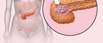
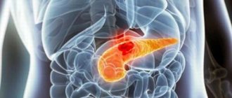
Pancreatic necrosis, or necrotizing pancreatitis, is a life-threatening complication of acute pancreatitis, which consists in the death of parts of the pancreas. The part “pancreo” translates to “pancreas,” and “necrosis” means “death.” Thus, the term “pancreatic necrosis” is redundant; just one word is enough.
Pancreatic necrosis occurs in 15-20% of patients with acute pancreatitis and occurs in moderate and severe forms.
Exacerbation
In the acute period, temperature indicates the activity of the inflammatory reaction. With the interstitial process, subfebrile numbers (37.5 ⁰C) are common. High temperature can occur only during moderate and severe processes in the iron. This is usually due to the release of enzymes into the surrounding tissues and an active inflammatory reaction. Fever in severe forms of the disease lasts about 3-4 days. Usually the numbers are above 38 ⁰C.
As mentioned above, temperature with pancreatitis is a poor prognostic sign. Typically, a pronounced febrile reaction indicates the extent of damage to the pancreas, the formation of infiltrates, and the formation of local or diffuse peritonitis.
Pancreatitis can occur without fever. This indicates a mild course or reduced reactivity of the body.
Classification of acute pancreatic necrosis
Classification of pancreatic necrosis is carried out on various grounds. Pathology can be classified depending on how much of the pancreas is involved in the process:
- An edematous form of pancreatic necrosis, in which the death of individual pancreatic cells occurs, without the formation of individual dead islets.
- Small focal pancreatic necrosis - affects up to 15% of the volume of the pancreas.
- Medium focal pancreatic necrosis - lesion volume up to 30%.
- Large focal pancreatic necrosis - up to 50% of the entire organ.
- Subtotal pancreatic necrosis - affects up to 75% of the pancreas.
- Total pancreatic necrosis - over 75% are exposed to necrosis.
Focal forms of pancreatic necrosis are called limited, and subtotal and total forms are called widespread.
Also, acute pancreatic necrosis is divided into infected and sterile, depending on whether infection is present in areas of dead tissue. Sterile pancreatic necrosis is in turn divided into:
- hemorrhagic;
- fatty;
- mixed.
Fatty pancreatic necrosis
A form such as fatty pancreatic necrosis develops slowly, about 4-5 days, which distinguishes it from the hemorrhagic form of sterile pancreatic necrosis. With fatty pancreatic necrosis, enzymes that break down fats - lipases - are more active. They destroy pancreatic cells and break down the internal fatty tissue of the organ.
With fatty pancreatic necrosis, a predominantly serous effusion is formed, the hemorrhages are small, and on the surface of the pancreas you can see plaques of steatonecrosis - foci of accumulation of calcium salts of fatty acids, which were formed due to the breakdown of fats. The fatty tissue that surrounds the pancreas is also subject to lipolytic damage.
Hemorrhagic pancreatic necrosis
Hemorrhagic pancreatic necrosis is characterized by a rapid course, pronounced intoxication of the body, and early onset of failure of many organs. With hemorrhagic pancreatic necrosis, an abundant bloody effusion is formed. There is total necrosis of the pancreatic tissue, purulent melting of individual acini - small structural units of the organ, massive bleeding into the pancreatic tissue.
Mixed pancreatic necrosis
Mixed pancreatic necrosis includes signs of both fatty and hemorrhagic pancreatic necrosis. It occurs most often, since during inflammation of the pancreas all mechanisms of damage to the tissues of the organ and adjacent structures work to an approximately equal extent. A clear division into hemorrhagic and fatty pancreatic necrosis is conditionally possible in laboratory conditions, during experiments when scientists artificially reproduce strictly defined factors of aggression.
Types of pancreatic necrosis
Pathogenetic mechanism
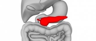
The pancreas is a parenchymal organ.
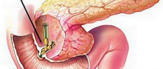
Its structure produces enzymes for digesting food. As they form, the secretion of the gland is formed - pancreatic juice. Through special ducts it enters the duodenum, where nutrients are processed. In the pancreas, enzymes are in an inactive state. They are fully activated upon contact with bile. The liver secretion, like pancreatic juice, enters the lumen of the small intestine through the sphincter of Oddi. This is a reflex mechanism that ensures timely activation of enzymes and uniform mixing of secretions with the food bolus.
When the pancreas becomes inflamed (pancreatitis), its parenchyma swells. This is accompanied by an increase in pressure inside the ducts and a violation of the outflow of pancreatic juice. Congestion and disturbances in the peristaltic activity of the ducts contribute to the reflux of bile into the pancreatic structure. Intraorgan activation of enzymes occurs. Inflamed gland cells cannot protect themselves on their own. The breakdown of protein and fat structures begins directly inside the organ. The changes are accompanied by:
- tissue death;
- distribution of processed products throughout the body;
- development of internal bleeding.
If damaged areas become infected, abscesses, accumulation of pus, ruptures of purulent capsules occur, and widespread peritonitis (inflammation of the abdominal wall) occurs.
When pancreas cells die, the body suffers from the action of activated enzymes, which, along with the blood flow, spread to other organs, tissue breakdown products, and toxins produced by bacteria.
The first to be damaged are the detoxifying organs – the liver and kidneys, followed by the heart and brain. In severe pancreatic necrosis, patients die from encephalitis and multiple injuries to internal organs.
ICD codes
Each disease has its own code in the International Classification of Diseases, 10th revision, abbreviated as ICD-10. Pancreatic necrosis in this classification corresponds to the code K86.8.1.
In the International Classification of Diseases 11th revision, which will come into force in participating countries only on January 1, 2022, pancreatic necrosis can presumably be coded with one of the following codes:
- DC35 – Certain diseases of the pancreas;
- DC3Y – Other specified diseases of the pancreas.
Currently, it is worth focusing on ICD-10. Despite the fact that there is less than a year and a half left before the transition to ICD-11, this transition will not be instantaneous, since it will require significant technical and technological adaptation, which means that it will not be possible to see the new code in life soon.
Treatment methods
In most cases, pancreatic necrosis is noticed at an early stage, then a special diet is prescribed along with a course of medications. For some time, a patient with a diseased pancreas may be put on a therapeutic fast to remove toxins. Antispasmodics, anti-enzyme drugs, immunostimulating and antibacterial agents are often prescribed. In the early stages of pancreatic necrosis, it is possible to avoid surgery, which is dangerous because it is difficult to know which areas are affected.
Medicines
To treat the pancreas, diuretics are used, and a local novocaine blockade is performed. To combat gland necrosis, antihistamines are used, and antispasmodics are administered intravenously in the presence of severe pain. A patient with a diseased pancreas receives insulin; if his blood glucose level increases, protease inhibitors are used. Patients take choleretic drugs if there are no gallstones.
Mineral waters containing alkali, cooling the pancreas, and fasting may be appropriate. If the disease was noticed in time, diagnosed and treated in a timely manner, then recovery and relief from the symptoms of necrosis is possible in 1-2 weeks. When the gland cannot be cured therapeutically, the patient is referred to a surgeon.
Find out how the pancreas is treated with medications.
- Buckwheat soup - recipes with photos. How to cook delicious buckwheat soup with broth or milk step by step
- How to treat ringworm in cats at home. Preparations and folk remedies for ringworm and weeping lichen
- Vaseline for eyelashes
Folk remedies
During an exacerbation of necrosis, the patient experiences severe pain. However, not only traditional medicine can cope with this. If you don’t want to stuff yourself with chemicals, try treating your pancreas with folk remedies. For this, plants and berries are used that can fight inflammation and reduce pain during necrosis. Let's find out how to treat your pancreas using natural remedies.
A decoction of Japanese Sophora will be useful. Pour a glass of boiling water over a spoonful of herbs and leave for several hours. The decoction should be drunk warm and consumed before meals. The course of Sophora to combat gland necrosis should not last more than ten days. It should take several weeks before using it again. The action of Sophora is primarily aimed at alleviating pain from pancreatic necrosis.
Blueberries are useful as an anti-inflammatory agent for necrosis. Leaves or berries are brewed, no matter dried or fresh. Use the drink as a daily drink to treat your pancreas. Immortelle has a similar effect. A spoonful of dry immortelle should be poured with a glass of boiling water. The resulting decoction, which must be divided into three times, should be enough for a day. This is a good remedy for the treatment of gland necrosis. Treatment of pancreas with folk remedies involves the use of oat decoction. It also relieves irritation and promotes the removal of toxins.
Causes of this pancreatic disease
The causes of the disease in most cases lie in alcohol abuse and poor diet. They account for 55% of all acute pancreatitis.
The second place is occupied by gallbladder pathologies. Stones can enter the common bile duct and the ampulla of the major duodenal papilla, which is common to the bile ducts and the main pancreatic duct. In turn, an increase in pressure in this system can lead to the reflux of bile into the pancreas, which causes inflammation. The share of these reasons is about 35%.
In 4% of cases, the cause is trauma to the pancreas. Most rarely, pancreatic necrosis occurs as a result of an autoimmune process and an infectious lesion.
Diagram of causes of pancreatic necrosis
Causes
The mechanism of necrosis is the inability of pancreatic tissue to resist the destructive effect of aggressive enzyme juice. Pancreatic juice has an alkaline reaction, which, after entering the intestine, is neutralized by the acidic contents of the stomach. But in cases where enzymes cannot be removed from the gland, alkali breaks down the protein elements of the cells. The destruction spreads to the blood vessels piercing the gland and creates injuries from which blood leaks. The process of destruction of organ cells by enzyme juice is called autoaggression.
According to the international classification of diseases, the pathology belongs to the subgroup “Acute pancreatitis” with code K85.
The more pancreatic juice is produced, the faster self-digestion occurs, and the more acute its manifestations.
Hemorrhagic pancreatic necrosis can cause damage and death of cells in other organs located in close proximity to the pancreas.
The following factors can provoke the onset of a pathological process:
- inflammatory foci in the gland that arise due to impaired excretion of enzyme juice;
- systematic toxicity with ethyl alcohol over a long period of time;
- retention of pancreatic juice in the ducts;
- infectious diseases of the biliary tract (cholecystitis, cholangitis, etc.);
- blockage of the bile ducts (with cholelithiasis);
- increased blood clotting in blood vessels, which accompanies malignant neoplasms, and thrombosis of blood vessels inside the organ after high doses of radiation;
- autoimmune disorders (vasculitis);
- complications after viruses and severe infections;
- overdose of certain groups of drugs;
- unbearable psychological stress;
- injuries and complications after surgical interventions on the organs of the food system.
The most aggressive enzymes contained in pancreatic secretions are produced to break down protein molecules of food entering the intestines. Elastase, trypsin and chemotrypsin lead to rapid destruction of gland parenchyma cells, sometimes affecting large areas. Because of this, the pancreas becomes inflamed and increases in size, which poses a significant threat to human health.
Predisposing factors for the appearance of this formidable disease are recognized:
- unhealthy diet with an abundance of fats and alcohol in the diet;
- pancreatitis in acute or chronic form;
- Constant consumption of trans fats.
Symptoms
Pancreatic necrosis always begins with acute pancreatitis. Symptoms often manifest themselves intensely: severe pain in the epigastrium, often of a girdling nature, and repeated vomiting that does not bring relief. At the very beginning, it is very important not to confuse pancreatic pain with fasting pain that occurs from hunger: food in the stomach will stimulate the activity of the pancreas, which will worsen the inflammation.
The more severe the attack of acute pancreatitis and the larger the volume of the pancreas is involved in the necrotic process, the brighter the symptoms, and the more rapidly the patient’s condition deteriorates, up to a critical drop in blood pressure, dehydration due to uncontrollable vomiting, unbearable pain, and loss of consciousness.
Conservative treatment of the disease
If the patient is not in a coma and conservative therapy is possible, then treatment begins with complete rest and fasting. Strict bed rest is prescribed, exercise (including walking), and eating are prohibited.
In the first 5–7 days, parenteral nutrition is prescribed with special nutrients, balanced in the composition of proteins, fats, and carbohydrates. The number of solutions administered and their composition are calculated individually, taking into account the severity of the patient and the degree of dehydration of the body.
In parallel, drug treatment is carried out aimed at:
- for pain relief to prevent pain shock;
- temporary blocking of pancreatic enzyme activity;
- decreased secretory function of the stomach and gallbladder;
- relieving spasms and improving the patency of the ducts;
- suppression of pathogenic microflora to prevent or treat infection or the development of toxemia;
- detoxification - removal of toxins that are formed during cell breakdown;
- detoxification;
- prevention of bleeding and suppression of secretion of the digestive organs by administering somatostatin - the growth hormone of the hypothalamus;
- preventing the development of infections.
Treatment algorithm
When a pancreatic necrosis clinic appears, it is necessary to urgently call an ambulance. Before the doctor arrives, you must be completely hungry, apply cold to the pancreas area and rest.
In case of severe pain, you can use an antispasmodic. It is not recommended to take painkillers before being examined by a specialist. The patient must lie down. The pain is relieved by lying on your side with your legs pulled up to your stomach.
If there are no indications for emergency surgery for pancreatic necrosis, the patient is placed in the intensive care unit, where intensive therapy is carried out for 3 days. In the absence of positive dynamics, the patient is transferred to the surgery department, where the issue of surgical intervention methods is decided.
Subsequently, rehabilitation is carried out, including medication and diet therapy, on which the patient’s condition and the speed of his recovery largely depend.
Medicines
Treatment with drugs for pancreatic necrosis is carried out in a hospital setting, often in the intensive care unit. Treatment tactics depend on the severity of the patient's condition. Therapy includes a set of measures aimed at restoring the functions of the digestive organs. For this purpose the following are used:
- antispasmodics, anticholinergics, analgesics (No-shpa, Papaverine, Platyfillin, Drotaverine, Baralgin, Ketanov) - for the purpose of pain relief;
- Novocaine blockade (Novocaine with glucose or Promedol, Atropine, Diphenhydramine is used);
- protease inhibitors (Contrical, Trasylol, Gordox, Pantripin) - to reduce enzyme activity;
- artificial somatotropin - growth hormone produced by the pituitary gland (Octreotsid, Sandostatin) - is used to inhibit the exogenous function of the organ; significantly reduces the area of necrosis; with prophylactic administration before surgical procedures, the production of enzymes by the gland is reduced;
- drugs from the PPI group (Pariet, Controloc, Omez), H2-histamine receptor blockers (Kvamatel, Cimetidine) - used to reduce the acidity of gastric juice, which stimulates the production of pancreatic enzymes;
- an antibiotic to treat or prevent infection (group of cephalosporins and fluoroquinolones) in combination with an antimicrobial drug (Metronidazole);
- solutions of electrolytes and protein blood products (Hemodez, Reopoliglyukin, Poliglyukin, plasma, albumin) - in order to combat shock, the infusion method of their administration is used;
- antihistamines (Diphenhydramine, Suprastin);
- diuretics (Lasix);
- antioxidants (Mexidol) - administered intramuscularly, intravenously or directly into the common pancreatic duct, prevent the formation of lipid peroxidation products (LPO). LPO causes irreversible changes in cells, leading to their death.
It is often necessary to perform therapeutic plasmapheresis and/or peritoneal dialysis.
How to diagnose
Diagnosis of pancreatic necrosis is not particularly difficult. First of all, an anamnesis is collected. Establishing the fact of long-term consumption of alcoholic beverages or cholelithiasis allows the doctor to draw the correct conclusion. Next, the surgeon conducts a physical examination and palpates the abdomen.
In a biochemical blood test in acute pancreatitis and pancreatic necrosis, characteristic changes are observed: an increase in the level of amylase and trypsin. In a general urine test, you can see the presence of diastase, which will also indicate damage to the pancreas.
An ultrasound examination of the internal organs is performed, after which a computed tomography scan of the abdominal organs is sometimes performed. CT is a more accurate and informative method, but at the same time more complex and expensive.
Types of disease and diagnostic methods
Hemorrhagic pancreatic necrosis is very dangerous and can cause death within 24 hours. Its common name is gangrene: tissues decompose and rot. It has been established that alcoholism does not always cause the development of this type of pancreatic necrosis. Often, a single use of alcohol in combination with fatty foods is enough for autolysis to suddenly develop - the flesh begins to rot and decompose all organs and systems, having a poisonous effect on them from the inside.
The cause of hemorrhagic pancreatic necrosis is the aggressive effect of pancreatic enzymes on its own tissues. Its development occurs under the influence of a large number of proteases - enzymes that break down proteins. One of them - elastase - acts on the walls of blood vessels, destroying them with the formation of hemorrhages.
Pathological anatomy: the organ is uniformly enlarged, edematous, there are many areas of necrosis in different parts of the gland - this is a large-focal type of pathology. Histology - swelling of the parenchyma with hemorrhages is observed.
If intensive therapy is not carried out in a timely manner, the organ may decompose completely due to the rapid breakdown of cells and tissues under the influence of an increasing amount of proteolytic enzymes.
Fatty pancreatic necrosis develops when lipase is synthesized in large quantities, destroying the adipose tissue of the gland. It develops gradually over several days. With timely provision of emergency care, the course is more favorable compared to the hemorrhagic process.
Mixed pancreatic necrosis is the most severe type of disease: all types of tissue are destroyed: protein, fat.
Diagnostics
Timely diagnosis is important if pancreatic necrosis is suspected. It is based:
- on complaints;
- medical history;
- laboratory research;
- functional methods.
Laboratory methods include blood tests:
- general clinical (leukocytosis, increased ESR, granularity of neutrophils, high hematocrit due to dehydration);
- biochemical (high enzyme content, positive C-reactive protein, aminotransferase levels exceeding the norm - ALT, AST);
- sugar (made repeatedly, dynamically).
Functional methods include:
- Ultrasound - ultrasound examination of the pancreas and abdominal organs;
- CT - computed tomography;
- MRI - magnetic resonance imaging;
- puncture of existing formations;
- diagnostic laparoscopy.
The first of these studies is ultrasound. The gastroenterologist may prescribe other additional methods and explain why they are necessary. Feedback from a specialist will help you navigate through numerous tests and examination methods.
Signs on ultrasound
Ultrasound examination of the abdominal organs is a simple and quick method for diagnosing pancreatic pathologies, which include acute pancreatitis and its complication – pancreatic necrosis.
First of all, the attention of the diagnostician will be attracted by an increase in the size of the pancreas and its swelling, indicating the active course of the inflammatory process. Ultrasound signs of pancreatic necrosis include dilation of the main pancreatic duct, caused by an increase in intraductal pressure due to blockage of the ducts by a stone or due to spasm of the sphincter of Oddi.
Signs on ultrasound
Main symptoms and complications
Long-term practice of studying and combating pancreatic necrosis allows us to conclude that its harmful attack on the body occurs, as a rule, very quickly.
For no obvious reason, the patient suddenly begins to feel heaviness in the abdomen and attacks of nausea, which transform into prolonged, debilitating vomiting.
With further development of the disease, acute pain appears in the left hypochondrium. Some symptoms may resemble a heart attack, but the doctor diagnoses that such signals are sent by the pancreas when pancreatic necrosis is located in the back.
Irradiation (spread of pain) under the scapula and into the left shoulder is also a characteristic sign of this disease.
Other symptoms characterizing pancreatic necrosis:
- Prolonged vomiting, without obvious relief.
- Increased temperature, chills, fever.
- The appearance of a painful skin color: paleness and redness of the skin.
- Intestinal paresis or paralysis is a neurological syndrome characterized by a lack of intestinal motor activity (peristalsis), as a result of which excrement is not excreted from the body.
- Increased heart rate, shortness of breath.
- Due to vomiting - dehydration of the body, drying of the mucous membrane in the mouth.
- The stomach swells, the muscles in its upper part tense.
- Urination decreases or completely stops.
- Around the navel, on the buttocks, and the costal arch on the back, characteristic bluish spots appear.
- General weakness sets in, or, as people say, weakness of the body.
- An imbalance in the patient's mental state manifests itself: unmotivated agitation, anxiety, confusion of thoughts, speech, consciousness, loss of spatio-temporal orientation, general lethargy.
- As a result of deep vascular damage, gastric and intestinal bleeding occurs.
Destructive changes associated with damage to the pancreas can provoke the following complications:
- The formation of voids filled with pus and necrotic masses, threatening the spread of an abscess.
- Development of pseudocysts and cysts in the body.
- The occurrence of fibrosis, as a result of which dead working cells are replaced by simple connective tissue, while the lost functional load is not restored.
- Limitation of pancreatic secretion – enzymatic deficiency.
- Acute purulent inflammation is phlegmon of the retroperitoneal tissue.
- The occurrence of thrombosis in the mesenteric vessels and portal vein.
The progressive development of pancreatic necrosis not only causes an increase in the size of the pancreas, but also leads to the formation of infiltrates - atypical compactions consisting of lymph, blood and dead cells. On the fifth day, the infiltrate is easily detected by palpation.
Clinical treatment guidelines
Conservative treatment, which does not involve the intervention of a surgical scalpel, is possible only for uncomplicated acute pancreatitis.
Conservative therapy
This therapy comes down to fasting in the first few days of the disease, complete rest and bed rest, and local cold on the stomach.
Patients with acute exudative (edema) pancreatitis are given intravenous infusions, painkillers, anti-inflammatory and antispasmodic drugs.
The occurrence of a complication in the form of pancreatic necrosis requires emergency surgical intervention.
Diet
The only diet that has a therapeutic effect in the acute phase of the disease is fasting, which lasts until the inflammation decreases to the point where food intake does not provoke new attacks.
The diet for pancreatic necrosis, when the life-threatening period is left behind, comes down to thermal and chemical sparing:
- food at a comfortable temperature is consumed, often stewed or boiled;
- Everything smoked, fried, salted, and an abundance of spices are excluded; many foods and alcohol are prohibited.
Insufficiency of insulin production, if it occurs after pancreatic necrosis, will lead to strict counting of bread units and calculation of the required amount of injectable insulin.
Operations
Surgical treatment of pancreatic necrosis involves removing dead parts of the pancreas and draining the abdominal cavity to remove effusion.
Surgery on the pancreas requires a lot of experience and a lot of luck from the surgeon: the organ is located deep in the body, it is difficult to get to it, the tissues of the gland are very delicate and difficult to operate on, and large vessels pass nearby.
The smaller the volume of intervention, the better for the patient, but the more pancreatic tissue is necrotic, the more traumatic the operation will be, and the worse the prognosis.
After surgery, many patients develop an external pancreatic fistula, which, after stopping the inflammatory process, is treated conservatively and closes on its own in an average of 2-4 months. If this does not happen, additional surgery may be required.
Surgery for pancreatic necrosis
Remission
In case of chronic inflammation of the pancreas, the thermometer should not show high numbers. Usually this period proceeds smoothly. A temperature above 38 ⁰C during remission indicates that secondary changes have formed in the pancreas (cysts, infiltrates, cavities) that have undergone suppuration. This process occurs after severe pancreatic necrosis.
What to do in this case? Contact your doctor immediately. Only he will determine what pathology can give such a reaction. With purulent inflammation against the background of pancreatic necrosis, in addition to the temperature criterion, other symptoms appear: pain, intoxication, bacterial changes in the blood (toxic granularity of neutrophils, shift of the leukocyte formula to the left). Abscesses and cysts are clearly visible during ultrasound diagnostics. What to do with them next is decided by the surgeon. Conservative treatment will not help here. Minimally invasive operations (puncture of the abscess under ultrasound guidance), laparoscopic techniques and open intervention are used.
Temperature during pancreatitis, especially during the chronic stage, is not always associated with the process in the pancreas. After illness, patients' immunity suffers greatly. Even a common respiratory virus can be severe with fever. Therefore, consultation with a competent doctor is so important for the patient. The doctor can notice the symptoms of developing complications and distinguish them from a simple cold.
Recovery period
Pancreatic necrosis is a serious complication of acute pancreatitis, which can be fatal and negatively affects many organs and systems of the body. It is logical that the recovery period after pancreatic necrosis can be up to a year or longer.
In terms of recovery, much depends on how much of the pancreas has undergone necrosis and self-digestion, how extensive the operation was, and whether the intervention was abdominal or whether everything was done using minimally invasive laparoscopic methods.
Due to pancreatic necrosis, people can spend entire weeks in intensive care and up to several months in the surgical department. Often patients lose a lot of weight. It may take several years to regain the lost pounds, since a strict diet does not provide the opportunity to eat up in a short time.
Questions and answers
Question: Are there any chances of getting pregnant and bearing a healthy child after an attack of pancreatic necrosis?
Answer: In most cases, doctors do not advise women to plan a pregnancy, as there is a high chance of fetal failure, miscarriage, early placental abruption or the birth of non-viable offspring.
Sometimes the development of severe toxicosis and exacerbation of diseases of internal organs serves as a reason for forced termination of pregnancy in the first trimester in order to save the woman’s life. But in medical practice, cases of the birth of healthy children from mothers who have suffered pancreatic necrosis have been recorded.
When deciding to take this step, a woman should:
- achieve stable (more than a year) remission of the disease;
- strictly adhere to the recommendations of specialists regarding nutrition and physical activity;
- realistically assess all the difficulties and dangers of possible consequences.
In some cases, it would be useful to consult a psychotherapist.
Question: Can one drink of alcohol lead to death several years after an attack of pancreatic necrosis?
Answer: Yes. And the combination of alcohol with fatty foods reduces the chances of survival by 3-4 times.
Question: What factors, besides diet, influence life expectancy after pancreatic necrosis?
Answer: In this case, excessive physical activity or, conversely, its complete absence can reduce life expectancy.
The stress factor plays a significant role. Constant psycho-emotional stress, overwork, lack of proper rest and sleep often leads to relapses of the pathology.
The following also have a negative effect on the body:
- development of a depressive state;
- loss of interest in life;
- apathy alternating with causeless irritability.
All factors, to one degree or another, influence how long they live after the development of pathology, as well as the quality of life itself. Prevention aimed at reducing the functional load on the pancreas helps avoid the development of the disease.
Consequences of the disease and possible complications
Subsequently, type 1 diabetes may develop if the remaining part of the pancreas is unable to produce the required amount of insulin. Chronic pancreatic enzyme deficiency is likely, requiring the use of enzyme preparations on a regular basis. Such consequences of pancreatic necrosis can and often do lead to disability.
Pancreatic necrosis is a very severe, life-threatening pathology. Complications of pancreatic necrosis include multiple organ failure and toxic shock.
Laboratory symptoms of pancreatic necrosis
The main diagnostic criterion for determining pancreatic necrosis is an increase in the level of amylase (an enzyme that breaks down starch) in the blood by at least 4 times. But if the patient was taken to the hospital not on the first day after the onset of the illness, but the next day, it is also advisable to determine the level of lipase (an enzyme that breaks down fats). It lasts longer. Acute pancreatitis is indicated by a 2-fold increase in lipase concentration. But even the detection of elevated levels of pancreatic enzymes in the blood does not indicate pancreatic necrosis. The amount of amylase in plasma may also increase with:
- necrosis of a section of the small intestine;
- ectopic pregnancy;
- intestinal obstruction;
- perforation of duodenal ulcer;
- diabetic ketoacidosis.
In the CIS countries, amylase levels are also determined in urine. Much less often the concentration of other pancreatic enzymes - elastase and trypsin - is detected in the blood plasma. And the method that determines the amount of P-type isoamylase is very rarely used. It is more specific, but has a high cost, so it is not practiced in all laboratories. Other, less significant laboratory signs of pancreatic necrosis:
- increase in the number of leukocytes in the blood;
- decrease in the amount of protein in plasma;
- increased blood glucose levels;
- incriminating the concentration of C-reactive protein;
- increasing the amount of procalcitonin (the study is expensive, so it is not used everywhere);
- an increase in the amount of liver enzymes in the blood (aspartic and alanine aminotransferases).
The above laboratory signs also do not provide 100% confidence in the diagnosis. They occur not only in pancreatic necrosis, but are also characteristic of myocardial infarction, viral and toxic hepatitis, and intestinal necrosis. Therefore, the diagnosis is made on the basis of instrumental examination methods.
Causes of pancreatic necrosis
Reasons that can provoke gland necrosis:
- excessive consumption of strong alcoholic drinks;
- binge eating;
- chronic diseases of the gastrointestinal tract and biliary tract;
- violation of dosage when taking certain medications;
- infectious diseases;
- stress and prolonged emotional tension.
Pancreatic necrosis, according to the scale of distribution, is divided into focal and extensive. Depending on a number of factors, the disease can be either sluggish or rapidly progress, involving neighboring organs in the process.
With proper conservative treatment of pancreatic necrosis, in most cases the patient makes a complete recovery. If for some reason the patient does not receive timely medical care, digestive enzymes begin to corrode the gland from the inside.
An abscess forms, in which pus enters the abdominal cavity, and the patient begins to experience acute peritonitis. In this case, immediate surgical intervention is required, otherwise sepsis cannot be avoided, which can cost the patient his life.
Often, extensive acute necrosis is observed in people who abuse alcohol.
Symptoms of pancreatic necrosis
Often, the necrotic process develops against the background of pancreatitis. Frequent signs of the disease at an early stage are pain of a girdling nature, spreading from the lower back to the shoulder blade, and reminiscent of the pain of a heart attack.
In this case, the patient begins to vomit profusely, the body temperature rises, bloating and flatulence, yellowness of the sclera are noted, the skin becomes pale or, conversely, too red. The main symptom of necrosis is the appearance of blue spots on the sides of the abdominal cavity.
On palpation, pain and tension in the anterior abdominal wall are noted. During a diagnostic study, fluid accumulation can be seen in the pericardium and pleural cavity.
As the disease develops, paresis, bloating, and weak peristaltic noises increase. Failure of the respiratory, cardiovascular, gastrointestinal, renal and hepatic systems develops.
Against the background of a disturbance in the respiratory system, pulmonary edema appears, accumulation of transudate in the pleural cavity; if the cardiovascular system is damaged, hypotension, threadlike pulse, and myocardial ischemia are noted.
Often the patient begins to experience mental disorders in the form of confusion and excessive agitation. Sometimes jaundice appears against the background of liver dysfunction.
Complications during the course of the disease can manifest themselves in the form of abscesses of the pancreas and retroperitoneal tissue, internal and external fistulas, peritonitis and bleeding.

