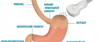
Information about the biliary system
Bile is a fluid that is produced in the liver. It helps digest food. Bile flows from the liver through the bile ducts into the small intestine.
If the bile duct is narrowed or blocked by scar tissue or a tumor, bile can no longer flow into the first part of the small intestine, the duodenum (see Figure 1). Therefore, bile accumulates in the liver. Stagnation of bile in the liver can cause infection, nausea, vomiting, fever, itching, and jaundice (the skin and whites of the eyes turn yellow).
Figure 1. Narrowed or blocked bile duct.
Biliary Drainage Catheter Information
If your bile duct is blocked, your doctor may suggest you have a catheter installed to drain the bile. This will help remove bile from the liver.
There are three different ways to ensure the flow of bile from the liver. Your doctor will discuss your options with you before the procedure.
- An external drainage catheter is inserted under the skin into the bile ducts. It is injected over the site of the blockage (see Figure 2). After this procedure, a catheter that comes out of your body will be attached to a drainage bag.
Figure 2. External biliary drainage catheter.
- An internal-external drainage catheter is inserted under the skin into the bile ducts and passed through the blockage. One end of the catheter will be inserted into the small intestine, and the other will be brought out of the body and attached to a drainage bag (see Figure 3). This catheter passes bile in two directions: either into an external container or into the small intestine. This is the most common type of drainage catheter, but this method is not suitable for all patients.
Figure 3. Internal-external biliary drainage catheter.
- During internal drainage (stenting) of the bile duct, a metal cylinder (called a stent) is placed to keep the blocked area open. After this procedure, a small catheter may come out of the body. If you have a catheter inserted, you will need to come back to the interventional radiology department later that day or the next day to have the doctor check that the stent is working properly. If everything is in order, the catheter will be removed.
The drainage catheter or stent is placed by an interventional radiologist. An interventional radiologist is a doctor who specializes in performing image-guided procedures.
After your procedure, during which you have a drainage catheter installed, it will be attached to a bile bag. Your doctor will tell you how much bile is expected to drain.
to come back to the beginning
Diet during rehabilitation
The gallbladder stored bile synthesized by the liver, and it was released into the small intestine. After the organ is removed, bile enters the intestines gradually and therefore it takes longer to digest food. To speed up digestion, it is important to follow dietary guidelines.
When eating the “right” foods, food will be digested faster, which means there will be no reflux, increased gas formation, fermentation and putrefaction in the intestines. In the first days you can only eat porridge, non-rich soups, fermented milk products, lean boiled meat, pureed vegetables, bananas. After discharge from the hospital, the patient must also adhere to a diet.
Recommended table No.5
It is forbidden to eat fried and fatty foods, smoked foods, spices, canned food, marinades, sweets, butter, eggs, and you cannot drink coffee or alcohol. Digestion will recover faster if you not only stick to a diet, but also follow a meal schedule. For a month after discharge from the hospital, it is necessary to organize 5-6 meals a day.
Before the procedure
Ask questions about your medications
You may need to stop taking some of your medications before the procedure. Talk to your doctor about which medications you can stop taking. Below are some common examples.
If you are taking medications that affect blood clotting, ask the doctor performing the procedure what is best for you. Doctor contact information is listed at the end of this material. These medications include:
| apixaban (Eliquis®) | dalteparin (Fragmin®) | meloxicam (Mobic®) | ticagrelor (Brilinta®); |
| aspirin | dipyridamole (Persantine®) | nonsteroidal anti-inflammatory drugs (NSAIDs), such as ibuprofen (Advil®) or naproxen (Aleve®) | tinzaparin (Innohep®) |
| celecoxib (Celebrex®) | edoxaban (Savaysa®) | pentoxifylline (Trental®); | warfarin (Coumadin®) |
| cilostazol (Pletal®) | enoxaparin (Lovenox®) | prasugrel (Effient®); | |
| clopidogrel (Plavix®) | Fondaparinux (Arixtra®) | rivaroxaban (Xarelto®); | |
| dabigatran (Pradaxa®) | heparin (subcutaneous administration) | sulfasalazine (Azulfidine®, Sulfazine®) |
Your doctor will decide whether to continue taking a certain medicine depending on the reason you are taking it. Do not stop taking any medications without talking to your doctor.
Review the resource Common Medicines Containing Aspirin and Other Nonsteroidal Anti-Inflammatory Drugs (NSAIDs). It contains important information about the medications you should not take before your procedure and what medications you can substitute.
If you take insulin or other medications for diabetes, you may need to change your dose before your procedure. Ask the doctor who prescribed your diabetes medication what you should do the morning of your procedure.
If you are taking any diuretics (medicines that make you urinate frequently), you may need to stop taking them on the day of your procedure. These drugs include furosemide (Lasix®) or hydrochlorothiazide. Talk to your doctor.
If you have ever been allergic to contrast dye, tell the doctor performing the bile duct drainage catheter procedure. He may prescribe medications for you before the procedure.
Let us know if you are sick
If you are feeling unwell (fever, cold, sore throat, or flu) before your procedure, call the interventional radiology nurse at 212-639-2236. Nurse working hours are Monday to Friday from 9:00 to 17:00. If you call after 5:00 p.m., on weekends, or on holidays, dial 212-639-2000 for the interventional radiology technician on call.
Record the time and place of your visit
Interventional Radiology will call you two business days before your procedure. You will be told what time you should arrive at the hospital for the procedure. If your procedure is scheduled for Monday, you will be called the previous Thursday.
You will also be told where to go on the day of your procedure. The operation will be carried out at one of the following addresses:
|
|
If you do not receive a call by 12:00 noon on the business day before your procedure, call 212-639-5051.
If for any reason you need to cancel your procedure, please notify the doctor who scheduled it.
Instructions for eating and drinking before your procedure
- Do not eat after midnight the night before your procedure. This also applies to hard candies and chewing gum.
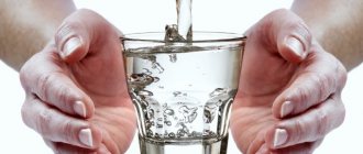
- Between midnight and two hours before you are scheduled to arrive at the hospital, you can drink no more than 12 ounces (350 ml) of water (see illustration).
- Avoid eating or drinking two hours before your scheduled arrival time at the hospital. This also applies to water.
to come back to the beginning
How is the drainage procedure carried out?
Drainage does not require preliminary preparation, so the manipulation is carried out according to the following algorithm:
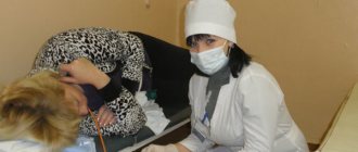
- Taking a general and biochemical blood test to study general indicators (coagulability must be checked);
- Before the operation, the patient is prescribed antibacterial therapy, since involuntary infection may occur during surgery;
- Drainage does not imply the administration of general anesthesia, since during the operation painkillers and sedatives are infused intravenously, and the patient is connected to equipment to monitor the general condition of vital organs;
- The surgical operation is performed using radiography with the introduction of a contrast agent, which allows you to monitor the overall progress of the surgical intervention;
- Direct drainage is based on the introduction of tubes into an area located slightly above the area of blockage of the bile duct and subsequent insertion into the organ. The tubes are brought out and sutured to the skin, and a container for collecting bile is connected to the system;
- After all necessary manipulations have been completed, the patient is transferred to the ward for further recovery.
When carrying out internal drainage, a special endoprosthesis is used, which is connected to the bile duct and the second end is connected to the intestine. The device is made of metal or polymer material.
If positive dynamics and favorable recovery of the body are observed, then the outer tube is subsequently removed.
Day of the procedure
What you need to remember
- Take only the medications your doctor prescribed to take on the morning of your procedure. Wash them down with a few small sips of water.
- Do not apply cream or Vaseline® to your skin. You can use deodorants or light lotions to moisturize your skin. Don't wear makeup on your eyes.
- Remove all jewelry, including body piercings.
- Leave all valuables, such as credit cards and jewelry, at home.
- If you wear contact lenses, wear glasses instead if possible. If you don't have glasses, bring a contact lens case with you to the hospital.
What to take with you
- List of medications you take
- Medicines taken for breathing problems (such as inhalers), medicines for chest pain, or both
- Glasses case or contact lens container
- Health care proxy form, if you have completed one
- If you use a CPAP or biphasic positive airway pressure (BiPAP) machine to sleep at night, take it with you if possible. If you are unable to bring one with you, we will provide one for you to use during your hospital stay.
What to Expect
When you arrive at the hospital, doctors, nurses, and other medical staff will ask you several times to say and spell your name and date of birth. This is necessary for your safety. People with the same or similar names can undergo the procedure on the same day.
You will see a doctor who will install a catheter for you. He will tell you about the procedure and ask you to sign an informed consent form.
You will be taken to the treatment room. If you do not have an intravenous (IV) line, your nurse will install one for you. You will be given sleeping pills through the IV catheter.
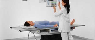
The catheter site will be numbed with an injection of anesthesia. To place the catheter, the doctor will use fluoroscopy (real-time X-ray).
to come back to the beginning
How is drainage performed?
The operation takes one and a half to two hours. No special patient preparation is required. Therefore, the intervention is carried out both planned and emergency.
External drainage of the bile ducts is carried out in a certain sequence:
- Blood is taken for analysis. In addition to the clinical one, a clotting test is carried out.
- On the day of surgery, the patient takes antibacterial agents. Infection may occur during surgery. It is important to prevent its development.
- The patient is not put to sleep during the operation. The patient is conscious. Through a venous catheter, sedatives and painkillers enter the bloodstream. Equipment is also connected to the patient to monitor blood pressure and other vital parameters.
- Surgery is performed in an operating room with X-ray equipment. This need is associated with ensuring control over the manipulations being carried out. For the same purpose, the patient is injected with a special solution – contrast.
- Using X-ray equipment, the doctor injects local anesthetics into the liver. An incision is then made.
- The next step is the introduction of external bile duct drainage. Using a guide, the tube is brought to a place located above the “blockage” area. Next, an “insertion” is made into the bile ducts. Tubes, the diameter of which is 2-3 millimeters, are brought out and sutured to the skin. The system is then connected to a special bag for collecting bile.
- After the operation, the patient is transferred to the ward.
Inoperable patients undergo internal drainage.
It requires an endoprosthesis. It is made of metal, polyethylene and other polymers. Metal endoprostheses last longer. The sequence of the internal drainage operation is similar to the external one. The exception is the installation of an endoprosthesis. The device is connected to the ducts along with a tube for external removal of bile. The other end of the endoprosthesis is brought out into the intestine. After carrying out control measures and identifying positive dynamics, the outer tube is removed.
After the procedure
After the procedure, you will be placed in a recovery room. You will need to stay in bed until the sedative medicine wears off.
In the recovery room, the nurse will continue to monitor your condition, heart rate, breathing and blood pressure. They will watch your catheter for possible bleeding.
About your catheter
A black mark will be placed on your catheter at the top of the disc (see Figure 4). The nurse will show it to you. This mark should always be at the same distance from the top of the disc. If the distance changes, the catheter has moved. You should call the interventional radiology department to have a staff member check the position of the catheter.
Figure 4. Black mark at the top of the disc.
The outer end of the catheter is connected to a 3-way stopcock (see Figure 5). It is called a 3-way stopcock because it has 3 connection points and a stopcock that can be turned to control the flow of bile. The drainage bag will be attached to the connection point opposite the catheter. The third connection point has a safety valve cap through which you can inject liquids. This valve is called a needleless connector.
Figure 5. Three-way valve.
A drainage bag will be attached to the catheter. You will see bile (a yellowish-green liquid) draining into the bag. The fluid may be blood red on the first day or the first two days. The color will eventually turn golden yellow or greenish depending on where the catheter is placed in your body.
The CathGrip® is a device that will prevent the catheter from leaving your body if you accidentally pull on the catheter.
to come back to the beginning
How long does rehabilitation last?
Depending on the cholecystectomy method chosen by the surgeon, the duration of rehabilitation will depend. After open surgery, ability to work is restored within 1–2 months, and after laparoscopy a person is on sick leave for no more than 20 days. The rehabilitation period can be divided into several stages:
Why does my stomach hurt when I lift something heavy?
- early (in hospital). Lasts two days after surgery. During this period, doctors monitor the patient’s condition: how he feels after anesthesia, and if there are any postoperative complications. If necessary, symptomatic therapy is carried out;
- late stage. The patient is still in the hospital. Doctors monitor how the body works without the gallbladder, whether intestinal function is disrupted, and how the wound is healing. This period is 3–6 days during laparoscopy and up to 14 days after laparotomy;
- outpatient rehabilitation. The body fully adapts to new conditions, digestion and well-being are restored. Depending on the type of intervention, it lasts 1–3 months.
For 4–6 hours after treatment, you are not allowed to eat or drink or get out of bed. After six hours you can get up, but only carefully, as you may feel dizzy after anesthesia. All patients experience pain in the incision area, but the intensity and duration of the pain varies.
So, after laparotomy, the patient is given narcotic painkillers (Promedol), and then non-narcotic analgesics (Tramadol, Paracetamol), and after laparoscopy, the pain is described as tolerable and the person does not need painkillers. The next day after removal of the organ, you are allowed to get up and walk around a little.
Upon discharge from the hospital, the doctor will tell you how to speed up recovery after cholecystectomy. Recommendations for diet, physical activity, medication, and suture care apply. Only by following the doctor’s instructions can you recover quickly and avoid postoperative complications.
Showering with a drainage catheter
You can shower, but your bandage must remain dry. Using a shower with a flexible hose will prevent water from getting on the bandage. You should also protect your adhesive bandage. If the adhesive bandage gets wet, change it. Wet bandages are a common cause of skin problems.
Before taking a shower, remove the belt and empty the drainage bag. Secure the drainage bag with tape to your body near the catheter, or use a belt to attach it to your waist. Use adhesive tape to secure cling film or a large enough plastic bag over the adhesive bandage to keep it dry.
You can also use AquaGuard, a disposable, waterproof coating to protect the adhesive bandage. When showering, make sure your catheter is completely covered to prevent it from getting wet.
Using the AquaGuard waterproof patch
- Figure 12. Folding the corners and removing the release tape from the AquaGuard patch
The AquaGuard waterproof patch has removable tape around the edges. Bend the corner of the tape on each side (see Figure 12).
- Hold the AquaGuard waterproof patch with the arrows facing up. Remove the top strip and attach the top edge of the AquaGuard waterproof patch over the adhesive bandage. Smooth it out.
- Then grab the folded corner and peel off the strip from one side, straightening the AquaGuard waterproof patch as it sticks.
- Follow the same steps for the bottom and other sides.
Do not allow the AquaGuard waterproof tape to touch the adhesive bandage. It may pull and rip off your bandage when you remove the AquaGuard waterproof patch after showering.
to come back to the beginning
Permitted activities with a biliary drainage catheter installed
- With the catheter in place, you can go to work and exercise. Avoid movements that involve lateral stretching or multiple bending. This may dislodge the catheter.
- Do not swim, bathe, or immerse the catheter in water. If you are planning a vacation, talk to your Interventional Radiology doctor about what to do in different situations.
- Make sure the tube is always securely attached to your body using the CathGrip.
- Do not jerk the catheter while getting dressed. Do not allow the tube to become kinked under clothing, such as tights or a belt. Try not to lie down on the catheter while sleeping. This will prevent the catheter from kinking. Typically, clothing hides the catheter.
to come back to the beginning
Drainage for obstructive jaundice
Obstructive jaundice develops against the background of improper flow of bile from the liver into the duodenum. As the disease progresses, bile acids and bilirubin enter the blood, which causes severe intoxication in the body.
In most cases, the cause of jaundice is considered to be the appearance of neoplasms in the gallbladder area:
- Tumors that are located near the bile ducts and compress the bile ducts;
- Metastases formed in the liver, which compress the organ and disrupt the natural flow of bile into the duodenum;
- Presence of stones in the ducts or gallbladder;
- Significant narrowing of the bile duct;
- Pancreatitis in acute or chronic form;
- Severe swelling of the pancreas;
- The appearance of formations directly in the bile ducts.
The formations described above are malignant in nature and in most cases are detected at the last stage of the disease, which provides irreversible consequences for the body.
The main symptoms of the disease: yellowness of the skin, whites of the eyes, mucous membranes, severe itching, aching pain in the epigastric region, white stool and dark urine.
Treatment of obstructive jaundice during tumor formation excludes the use of chemotherapy, so temporary drainage is necessary, which ensures normal outflow of bile and prevents the development of additional complications in the body.
Special instructions for internal-external biliary drainage catheter
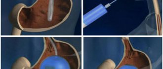
If you have an internal-external biliary drainage catheter, your doctor may suggest that you undergo a plug test. During this test, the outside of the catheter will be closed so that all the bile can flow down the catheter directly into your body and so that you no longer have to use a drainage bag (see Figure 13).
Figure 13. Closed biliary drainage catheter
Your catheter may be closed at the hospital before you go home, but you will most likely be discharged from hospital first and advised to close it after you have been home for a few days.
To close the catheter, remove the three-way stopcock from the catheter and attach a needle-free connector to the end instead.
Continue flushing the catheter on the same schedule through the needleless connector.
When the drainage catheter is closed
If you have closed your internal drainage catheter, you should watch for the following symptoms:
- leakage at the catheter site;
- pain in the abdomen (belly) around the catheter insertion site;
- temperature 100.4°F (38°C) or higher or chills.
These symptoms may occur any time after the catheter is closed. If you experience any of these symptoms, call and report it to Interventional Radiology. Then open the catheter. To do this, remove the needleless connector (cap) from the catheter and reattach the catheter to the drainage bag. Symptoms should disappear within 30 to 45 minutes.
Do not re-close the catheter without calling Interventional Radiology.
to come back to the beginning
Possible complications
Obstructive jaundice is a rather dangerous disorder. In the absence of timely treatment, severe pathological phenomena develop. Therefore, it is necessary to restore the patency of the bile ducts, improve the outflow of substances, and reduce the load on the organs.
Installing drainage is the most effective method of treatment. Unlike conservative methods, it allows you to quickly eliminate obstruction and restore normal outflow. External and external internal drainage is installed without complications. Such options are considered safe and in rare cases cause side effects. Most often, complications arise with the percutaneous transhepatic method.
This is interesting: How does the color of urine and feces change during jaundice? Causes of changes, treatment and prognosis
Negative consequences of the procedure:
- Hemorrhagic disorders
- Peritonitis due to leakage of bile into the abdominal cavity
- Pain syndrome
- Development of cholangitis
- Displacement of drainage catheter, tube, prosthesis
- Suppuration in the area of implementation
Thanks to the use of modern medical instruments and equipment, the risk of complications is minimal.
Call your Interventional Radiology nurse or doctor if:
- you have a temperature of 100.4°F (38°C) or higher;
- symptoms of obstruction (blockage), such as pain or leakage from the catheter;
- there is no outflow of bile through the catheter or its amount is much less than usual;
- the drained fluid resembles blood;
- leakage was detected around the installation site;
- it is impossible to flush the catheter, or leakage is detected during flushing;
- the position of the black mark has changed;
- there is no outflow of fluid from the catheter when the three-way valve is in a vertical position;
- the clamp is loose or open;
- there is a kink in the tube causing a blockage that you cannot straighten;
- the catheter insertion site is painful, tender to the touch, or swollen;
- the skin around the catheter is red, irritated, or different from its normal state;
- you feel sick;
- jaundice appeared or worsened;
- you are concerned about something about your catheter.
to come back to the beginning
Indications for drainage of the bile ducts
Drainage of the common bile duct has its own indications and contraindications. So, start draining if necessary:
Drainage of the common bile duct has its own indications and contraindications
- Remove infected and stagnant fluid from the liver and passages.
- Eliminate narrowing of the lumen of the common bile duct and restore the passage of bile to other organs.
- Rinse the gallbladder and its passages with antiseptics or antibiotics.
- To avoid scar formation at the surgical site in the gastrointestinal tract.
- To prevent the formation of fistulas after surgery on the duct.
- To drain fluid from the liver to clean the bladder cavity.
Order information
Before you go home, your nurse will provide you with enough supplies for 2 weeks of use. Usually, the nursing service will then order the materials for you.
If you would like to order your own supplies, please call Interventional Radiology at 212-639-2236 and place your order using the item numbers below. You can pick up your order in two business days at the IR Clinic located at 16 East 60th Street, between Fifth Avenue and Madison Avenue.
| Name | MSK center number | Delivery terms |
| Adhesive remover | 3170 | package |
| alcohol wipes; | 3330 | package |
| CathGrip | 2675 | PC. |
| Drain collection kit, sufficient for 4 weeks | 3115 | kit |
| Needleless connector | 9697 | PC. |
| Micropore Paper Plaster | 4326 | package |
| Non-sterile latex gloves | 4545 | package |
| Regular gauze | 3424 | package |
| Leather treatment solution | 3332 | package |
| Perforated dressing material Telfa | 3327 | PC. |
| Three-way tap (TM Cook) | 5192 | PC. |
| Uresil drainage bag | 3361 | PC. |
| Adhesive bandage Uresil | 3226 | package |
Pre-filled syringes
You will need a prescription for pre-filled syringes with normal saline. You can get a prescription from the IR Clinic. If your local pharmacy does not carry syringes, you can get them from the outpatient pharmacy at Memorial Hospital at 1275 York Avenue, between East 67th Street and East 68th Street. It is open from Monday to Friday, from 09:00 to 17:45. Pharmacy phone number: 646-888-0730.
to come back to the beginning