- February 1, 2019
- Tests and diagnostics
- Konstantin Kim
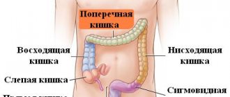
The large intestine is located in the pelvis and partly in the abdominal cavity, where it originates from the small intestine. It is the final stage of digestion and ends in the rectum.
The length of the large intestine reaches 1.5 m, the small intestine – up to 4 m. A bolus of food makes its full journey through the body in 14-20 hours. The small and large intestines differ in their structure and functions.
Bowel functions
The small intestine absorbs all incoming nutrients. Thick - absorbs water and forms feces, but its role does not end there. 2/3 of its contents are microflora, numbering up to 500 species of bacteria. In weight terms it is about 2 kg. Thanks to beneficial intestinal bacteria, the body is saturated with vitamins and enzymes, food is digested, a set of amino acids, hormones, and immunity is preserved.
If the diet is not followed, conditions are created in the intestines for putrefactive fermentation of foods and toxins are released. Rotting dietary fiber creates an alkaline environment that increases the growth of pathogenic bacteria.
The abdominal cavity contains many vessels and blood circulates intensely. The required temperature is set by the intestines not only for the occurrence of biochemical processes, but also for the regulation of heat throughout the body.
Stimulation of the work of internal organs lies in the fact that different parts of the large intestine contain biologically active points, massaging of which causes the internal organs to function intensively. This happens because they are projected onto the walls of the colon. Improved performance occurs due to increased blood flow and metabolism.
Removing waste feces is the most important condition for maintaining health. With constipation, these processes are disrupted and toxins begin to be absorbed into the body. Constant poisoning of the body leads to varicose veins, colitis, the growth of polyps, and cancer. Pathology can manifest itself in the form of flatulence, abdominal pain, the appearance of mucus and traces of blood in the stool, frequent changes in states of diarrhea and constipation.
Primary signs of bowel cancer
Like many other oncological diseases, intestinal cancer in the initial stages manifests itself with subtle and subtle symptoms, which often go unnoticed:
- certain changes in intestinal motility;
- general weakness, fatigue, loss of body weight that does not have natural causes;
- anemia;
- the presence of blood in the stool;
- changes in taste preferences, the appearance of aversion to certain foods.
The presence of such signs and symptoms of bowel cancer, as a rule, does not cause concern for patients. This oncopathology in the early stages can be detected by chance, when the patient undergoes an endoscopic examination for a completely different reason. Most often, intestinal cancer is detected in late, advanced stages, requiring long-term and complex treatment.
Early diagnosis of colon cancer ensures timely initiation of treatment, which increases the patient’s chances of recovery and maintaining quality of life. Therefore, if you have at least one of the above signs of this disease, you must immediately consult a doctor and undergo a thorough examination.
Diagnosis and treatment of oncological diseases is carried out in one of the leading centers in Moscow - the Yusupov Hospital.
Indications for bowel examination
Common symptoms of intestinal disorders include:
- stool instability for a long time;
- hardening of feces;
- pain in the anus and abdomen;
- flatulence;
- nausea and vomiting;
- bad breath;
- belching;
- the presence of blood and mucus in the stool.
Nonspecific symptoms may be added: malaise, weakness, signs of general intoxication.
Regular examination of the gastrointestinal tract and intestinal checks should be carried out for those who have already undergone surgery for a tumor in the intestine and for patients over 40. If there are patients in the family with diagnoses of intestinal oncology, Crohn's disease, polyposis, then the person is at risk.
Physical examination techniques
Physical techniques are studies that a doctor can conduct without the use of special equipment. These include a general examination of the abdomen, palpation and rectal examination. In rare cases, percussion and auscultation are used.
Examination of the patient
An examination is a visual assessment of the condition of the patient’s anterior abdominal wall. Using this technique you can evaluate:
- skin color, presence of pallor, cyanosis or local redness;
- elasticity, skin turgor;
- increased muscle tone of the abdominal wall is one of the symptoms of peritonitis;
- the presence of skin rashes, roughness of the integument.
An indirect symptom of disorders in the intestines can also be a varnished surface of the tongue or the appearance of a white coating on its surface.
Be sure to read: Preparation and performance of intestinal irrigoscopy
Palpation of the abdomen
Palpation is palpation of the abdomen, assessing the condition of the anterior abdominal wall. Depending on the depth of pressure, superficial and deep palpation are detected.
First, the patient undergoes superficial palpation of the abdomen. The doctor consistently presses on the surface of the abdominal wall, moving in a circle. The patient must relax the abdominal muscles. Signs of dysfunction of the digestive system, which can be detected by palpation, are:
- increased or weakened intestinal motility (strength of muscle contractions of the organ);
- the presence of pain limited to a certain area or spread over the entire surface of the abdomen;
- increased tone of the abdominal muscles - rigidity, indicating the spread of the process to the peritoneum.
In the place where pathological changes are determined, the doctor performs deep palpation. It involves pressing the fingers firmly into the abdominal wall, while the patient must exhale. When using deep palpation, the doctor can more accurately assess changes characteristic of diseases of the digestive system.
Rectal examination
A rectal examination is palpation of the anus and anal canal in order to assess their functional activity. The examination is carried out with the patient lying on his side with his legs drawn to his body and his knees bent.
A rectal examination is performed if the patient has local symptoms (itching and burning in the anal area, bleeding). Diagnosis is the first step in identifying such a common disease as hemorrhoids.
Read on the topic: Using a rapid test from a pharmacy to determine occult blood in stool
Bowel examination methods
Any examination begins with an external examination, questioning and history taking. After this, the proctologist must perform a digital examination of the rectal intestines. In this case, the doctor can assess the tone of the anus muscles, palpate possible diseases of the pelvic organs (scars, hemorrhoids, fissures, polyps, narrowing of the intestinal lumen, tumors).
Deeper sections are examined instrumentally. Such a study of the body allows the doctor to choose the next type of examination.
Modern methods of intestinal examination
There are a number of the most popular methods used to diagnose an organ.
Among them are the following:
- capsule examination;
- endoscopy;
- colonoscopy;
- irrigoscopy.
Let's look at each of them in more detail so that you can understand what kind of medical procedure a doctor can send you for in a given case.
Capsule examination
It is believed that this is the least invasive method, that is, safe in terms of infection. With its help, you can examine every part of the gastrointestinal tract of an adult. For this, a special enterocapsule with a small chamber is used.
Reasons for conducting such a survey:
- Unexplained pain in the abdominal area;
- Presence of hidden bleeding;
- Detection of signs of congenital pathology or tumor;
- Detection of symptoms characteristic of a malignant neoplasm.
The procedure for examining an organ using the capsule method is as follows: the patient comes for an appointment in the morning without having breakfast beforehand. A recording device is attached to his body and he is given a capsule with a camera to swallow.
Once in the stomach, it moves along it and passes into subsequent sections of the gastrointestinal tract due to waves of peristalsis.
A computer program analyzes the information received, and after eight hours the doctor receives the result. Having done its job, the capsule naturally leaves the body.
The main advantage of the capsule diagnostic method is convenience. During the examination, a person can lead a normal lifestyle and not limit himself in anything. In addition, this method, developed by Israeli specialists, is recognized throughout the world as the most highly informative method, capable of identifying even those pathologies that are hidden, for example, tumors and polyps.
Since a standard capsule moves through organs using peristalsis, this is impossible with minor wave-like contractions. In such cases, another type of capsule is used, which is called Patency. It is equipped with a microchip, not a camera.
If the capsule does not pass further through the gastrointestinal tract, it dissolves within a few days, and the chip informs the doctor where there is a pathological narrowing of the organ. Subsequently, the chip also comes out naturally.
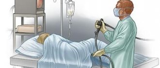
Endoscopy
Endoscopy is used to examine the body for the presence of polyps or tumors. In this case, the patient must adhere to certain rules of preparation for the procedure, in particular, cleanse the body in any convenient way (naturally, using a laxative or enema).
To carry out the procedure, a specialist inserts an ultrasound sensor into the rectum. At the location of the pathology, the recording device informs the doctor of the information received, namely the degree of spread of the resulting tumor.
The advantages of endoscopy are safety and painlessness, as well as the ability to obtain reliable data in the shortest possible time. The device allows you to visually examine the condition of not only the intestinal mucosa, but also the esophagus, stomach and duodenum.
Among the contraindications, which in general are not many for endoscopy, it is worth mentioning diseases in the area of organs such as the lungs and heart. This is due to the fact that the patient is taking special medications that may distort the examination data.
Colonoscopy
This method is a more unpleasant procedure, during which the patient may experience some discomfort, in particular minor pain and bloating.
The examination is carried out using a fiber colonoscope - a flexible tourniquet equipped with an optical system. In the process, you can not only visually examine the walls of the organ, but also perform a biopsy of presumably pathological tissue for histology. In addition, if there are small polyps or benign tumors, they can be removed without surgical methods.
By agreeing to a colonoscopy, the patient immediately after the examination receives highly accurate diagnostic data regarding adhesions, tuberculosis or tumors.
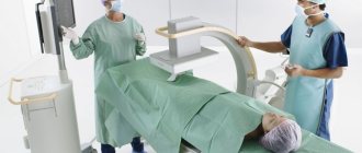
Irrigoscopy
Preliminary measures include bowel cleansing with laxatives or an enema, as well as abstaining from eating on the eve of the examination.
To perform irrigoscopy, the patient drinks a liquid with a concentrated radiopaque substance (most often barium sulfate), which, passing through the organs of the gastrointestinal tract, floods its areas.
Pictures taken by the doctor after this clearly show the contours of the intestine and the level of its lumen. The data obtained allows us to establish the presence of pathologies and the cause of their occurrence.
The irrigoscopy method is quite safe, since the degree of radiation exposure is very small, and is absolutely painless. It allows you to determine the presence of hidden voids, sac-like protrusions, tumors, developmental anomalies existing from birth, ulcers and scars by the relief of the shell.
Reasons for performing irrigoscopy:
- Discharge from the intestines of a mucous and purulent nature;
- Varying intensity of bleeding detected during bowel movements;
- Pain inside or outside, in the anus;
- Chronic diarrhea or, conversely, constipation;
- Inability to use colonoscopy for any reason;
- Suspicion of a tumor;
- Suspicion of a disease such as intestinal obstruction.
The results obtained using this diagnostic method can be confirmed using x-rays or ultrasound.
Methods for examining the large intestine
To diagnose the intestines for certain diseases, one method is not enough - an integrated approach is required.
First of all, the doctor must give a referral for a blood test - clinical and biochemical, as well as stool. In addition, a whole arsenal of techniques comes into play, including sigmoidoscopy, fibrocolonoscopy and anoscopy.
If there is a suspicion of diseases of the pelvic organs, a mandatory condition is to conduct a digital examination through the rectal opening using special instruments. To analyze problems in deeper sections, a device such as a proctoscope is used. Fibercolonoscopy is used to examine the entire organ.
Diagnosis of the small intestine
When diagnosing the area of the small intestine, research begins with its main components, namely the duodenum, ileum and jejunum, which are located in the space between the large intestine and the stomach.
Diagnostics
The method of examining the large intestine can be either instrumental or non-instrumental examination.
For the rectal area, 5 methods are used:
- primary digital examination;
- anoscopy;
- sigmoidoscopy;
- fibrocolonoscopy;
- laboratory diagnostics of stool for dysbacteriosis;
- blood analysis.
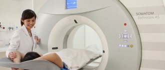
For a general complete examination of the colon, ultrasound, colonoscopy, MRI, CT, capsule ingestion, X-ray examination, irrigoscopy, and angiography are also used.
How to check the small intestine: methods
Given the location of the small intestine, it can be argued that access to it is usually difficult. Therefore, the condition of this organ is assessed in 2 ways. The first (FGDS) involves examining the organ through the oral cavity. This way you can see the initial part of the small intestine. The second diagnostic method is colonoscopy. In this case, visualization is carried out by inserting an endoscope through the anus. Colonoscopy can evaluate the condition of the distal small intestine.
In addition to endoscopic methods, there are other diagnostic methods. How to check the small intestine without colonoscopy and FGDS? The following methods of organ research are distinguished:
- Physical examination. It is the first stage in diagnosing diseases of the small intestine. Physical examination means palpation and percussion of the abdominal area.
- Laboratory research. Thanks to the tests, it is possible to find out whether there is an inflammatory process, as well as oncological pathologies. Laboratory diagnostic methods include: CBC, stool examination, cytology smear.
- X-ray of the abdominal organs with contrast. This method helps to identify the presence of changes in the intestinal walls, shadows from neoplasms.
- Biopsy and histological examination. Performed if an oncological process is suspected.
All of the listed diagnostic procedures are of great importance in identifying pathologies of the small intestine. Often it is necessary to perform several research methods.
Where does intestinal diagnostics begin?
Examination methods involve a primary examination of the small intestine - duodenum, jejunum and the beginning of the colon - ileum. A gastroenterologist deals with these organs.
For diagnostics use:
- fiberoscopy;
- irrigoscopy;
- endoscopy;
- ultrasonography;
- X-ray.
There are a lot of ways to study the intestines; they can be combined into radiation and non-radiation; endoscopic and non-endoscopic. Radiation methods are CT, X-ray, irrigoscopy. They are not indicated for children, pregnant women and women during lactation.
How to check the intestines in other ways?
There are various ways and methods in which you can conduct a bowel examination without a colonoscopy. Conventionally, they can be divided into invasive and non-invasive.
The first analogues include:
- Finger method of intestinal examination;
- Irrigoscopy;
- Anoscopy;
- Recotoromanoscopy;
- Capsule diagnostics.
The essence of each of these examinations is to examine the intestines from the inside using various devices, tubes, endoscopes and other things.
Examining the intestines using any of these methods will be less painful than using a colonoscopy, but discomfort will still be felt.
Non-invasive methods include:
- Ultrasound examination (ultrasound);
- Computed tomography (CT);
- Magnetic resonance imaging (MRI);
- Virtual colonoscopy;
- Endorectal ultrasound;
- Positron emission tomography.
When performing any of this list of intestinal examinations, the patient will not feel pain or unpleasant consequences of the procedure. However, such a check is not an alternative to colonoscopy, but only a possible addition .
The fact is that colonoscopy shows the presence of a tumor even at an early stage, reveals fissures and fistulas and is a more informative diagnostic test. And its main advantage is the ability to take a biopsy for oncology and remove various polyps and anomalies.
Therefore, you should not try to replace colonoscopy with any other methods of examining adults and children. It is better to supplement it than to explore it with other methods.
FEGDS
Endoscopic examination of the intestines allows you to check the condition of the esophagus, stomach and duodenum. It is carried out not only for diagnostic, but also for therapeutic purposes. During this method, the foreign body is removed, bleeding is stopped, and a biopsy is performed. This procedure is not performed only for heart or lung diseases. FEGDS is divided into planned and urgent.
Its advantages:
- availability;
- rapidity;
- good tolerability;
- information content;
- low invasiveness;
- possibility of carrying out on an outpatient basis.
The downside is discomfort when inserting the probe.
Indications for FEGDS:
- ulcer;
- gastroduodenitis;
- bleeding;
- cancer of the papilla of Vater;
- esophagogastric reflux.
Preparation for the procedure:
- The last 2-3 days before the procedure, diet with the exception of spicy foods, nuts, seeds, chocolate, coffee and alcohol.
- Dinner the day before no later than 18:00.
- Do not have breakfast or brush your teeth in the morning.
The patient is examined by laying him on his left side, with his knees pressed to his body. Local anesthesia in the form of lidocaine spraying. A thin tube with a camera is then inserted into the mouth.
At the doctor's command, swallowing movements are performed. An image of the parts of the intestine being examined is displayed on the monitor screen. Eating after the end of the manipulation is allowed after 2 hours.
Contraindications to FEGDS:
- rachiocampsis;
- mediastinal tumors;
- goiter;
- hemophilia;
- cirrhosis;
- esophageal stenosis;
- history of heart attacks and strokes;
- asthma with exacerbation.
Relative limitations are severe hypertension, lymphadenopathy, tonsillitis, laryngitis and pharyngitis, psychosis.
Methods for diagnosing the small intestine
Depending on what disease the doctor suspects, different diagnostic methods are used.
Radiography
A special option is used - irrigoscopy, when the patient drinks a contrast mixture, then a series of x-rays are taken. The contrast, moving along the digestive canal, outlines all folds, ulcers, polyps, tumors, diverticula (blind protrusions) and other formations. All parts of the intestine are examined at once.
Double balloon enteroscopy
It is performed under anesthesia due to the fact that inflation with gas causes pain. The endoscope is inserted through the mouth, the balloons are sequentially inflated: the near one is filled, and the far one is deflated, then vice versa. Gradually, the probe is advanced and the balloons are inflated until the entire intestine has been examined. At the same time, you can remove the polyp, stop the bleeding, or perform another manipulation.
Be sure to read: Sphincter of Oddi: characteristics and diseases of the anatomical structure
Ultrasound of the small intestine
A harmless and safe examination, accumulations of pathological fluid, tumors, enlarged lymph nodes, adhesions, cysts, developmental defects, fistulas, hematomas, and accumulations of parasites are visible. An important advantage is that you can observe the work of the intestines and glands in real time.
Hydro-MRI of the small intestine
Two types of contrast are used: oral, which is given to drink through the mouth, and intravenous, which is administered through a subclavian catheter. After the patient drinks the contrast, a series of images are taken, then they are repeated after intravenous contrast. Contrast agents accumulate in the walls of the small intestine. If there are contraindications ─ metal implants in the ear canal, rods in the bones or pacemakers ─ CT with the same principles of contrast is used. Almost all diseases are detected, including cancer metastases to the mesenteric lymph nodes.
Capsule endoscopy
The patient swallows an endoscopic capsule, which resembles a tablet in shape and size. The capsule has a built-in video camera and illumination device, as well as a radio signal transmitter. The receiving antenna and storage device are attached to the patient's body. The endoscopic capsule passes the entire digestive canal and exits naturally, taking many detailed pictures. Today this is the most informative examination method.
FGDS with biopsy
Main article: Fibrogastroduodenoscopy (FGDS): preparation and implementation
An endoscopic examination method that thoroughly examines the stomach, the place of transition to the intestines and the duodenum. Examination of deeper parts of the body using this method is impossible.
Colonoscopy
The main method for diagnosing colon diseases in women and men is intestinal colonoscopy. It is considered the most effective. During the procedure, a flexible probe with a camera at the end is inserted into the intestines through the anus. This method of checking the intestines allows you to visualize ulcers, polyps, inflammations, tumors, etc. During the examination, a biopsy, removal of polyps and foreign bodies, stopping bleeding, and restoring intestinal patency are possible. The main advantage of the innovation is the study of the entire large intestine.
The following indications exist for intestinal colonoscopy:
- neoplasms on the mucous membrane;
- inflammation and swelling;
- ulcerative colitis;
- Crohn's disease;
- presence of a foreign body in the intestine;
- intestinal obstruction and constipation;
- polyps;
- blood in stool, etc.
The insertion probe is 1.6 m long, soft and flexible, and has a video camera at its end. Local anesthesia is administered intravenously or applied to the endoscope to reduce pain during insertion. General anesthesia is recommended for children under 12 years of age.
Contraindications for colonoscopy:
- pulmonary heart failure;
- peritonitis;
- severe colitis;
- myocardial infarction;
- stroke;
- pregnancy.
Cleansing the intestines before the study is the main condition of preparation. This can be achieved by enemas or special laxatives. Castor oil can be used as an additional remedy for severe constipation.
2-3 days before the examination they adhere to a light, slag-free diet. It excludes:
- sources of fiber - greens, vegetables and fruits;
- smoked meats, pickles, marinades, rye bread, chocolate, peanuts, chips, seeds, milk and coffee.
To cleanse the intestines, after consultation with a doctor, you can use Lavacol, Endofalk, Fortrans, Duphalac. The latter is good because it does not irritate the mucous membrane.
Colonoscopy is an unpleasant procedure; insertion of the probe is painful. During the camera examination, the intestine is dilated by injecting air into a balloon for better examination.
This intestinal examination lasts 20-30 minutes. If the approach is incorrect, complications may arise in the form of bleeding, perforation of the wall, and bloating.
Instrumental diagnostic methods
Instrumental diagnostics is an examination of the intestines using specialized equipment. This group includes radiation research methods (x-ray, MRI, ultrasound) and endoscopic diagnostics (sigmoidoscopy, colonoscopy).
Colonoscopy
Colonoscopy is an examination of the large intestine using special endoscopic equipment that contains a camera and lighting elements. When the endoscope passes through the intestine, it “removes” the intestinal wall, and the resulting image is transmitted to the monitor. Thanks to this, the doctor can assess the nature of changes in the intestinal mucosa and detect pathological changes:
- tumors;
- cysts;
- inflammatory processes;
- polyps;
- areas of ulceration;
- local hemorrhages.
Some therapeutic manipulations can also be done directly during the diagnostic procedure. So, with the help of an endoscope, bleeding is stopped or polyps are excised. From suspicious areas of the intestine, you can take a biopsy - a fragment of the mucosal surface.
The biological material is sent for histological examination, where its cellular composition is determined.
Be sure to read:
MRI of the intestine and colonoscopy: which study is better?
A biopsy is performed in patients with suspected malignancy. This technique allows you to accurately confirm the nature of the tumor and carry out differential diagnosis with benign changes in the mucosa.
X-ray examination
X-ray examination of the intestines is divided into main types - radiography and irrigoscopy. Plain radiography has little diagnostic value, since it cannot visualize intestinal loops. However, the image will show severe disturbances in the digestive system:
- places where fluid accumulates;
- the appearance of gas in the abdominal cavity (may indicate intestinal perforation);
- intestinal obstruction, which is characterized by the accumulation of the contents of the digestive system in one area and the lack of movement of food masses.
Irrigoscopy
Irrigoscopy is of greater value in diagnosis. The technique involves introducing a contrast agent into the patient’s gastrointestinal tract - a composition that is clearly visible on an x-ray. By the nature of the distribution of contrast in the digestive system and the speed of its passage through the stomach and intestines, one can judge the functional activity of the organ.
Irrigoscopy allows you to detect:
- pathological changes in the walls of organs (ulcers, tumors and polyps protruding into the intestinal lumen);
- narrowing of intestinal loops;
- the presence of accumulations of food masses (intestinal obstruction);
- inflammatory changes on the surface of the mucosa.
Irrigoscopy is an important component in diagnosing diseases of the gastrointestinal tract.
Ultrasound diagnostics
Ultrasound is an examination technique that is based on the reflection of ultrasound rays from the organs of the digestive system and their recording by a special sensor. The technique is completely safe for patients and can be used if there are contraindications to radiation exposure.
Using ultrasound, you can evaluate the size and shape of intestinal loops, the thickness of the mucosa, and the structure of the organ wall. On the screen you can detect signs of pathological changes (tumors, inflammatory processes). In addition, ultrasound is used to examine the liver and pancreas, with diseases of which it is necessary to differentiate disorders in the intestines.
CT scan
Computed tomography (CT) is a highly sensitive diagnostic technique that can even obtain layer-by-layer images of the abdominal organs.
Tomography allows you to identify signs of the following diseases:
- polyps and other benign neoplasms;
- malignant tumors;
- inflammatory diseases;
- intestinal bleeding.
The high accuracy of images makes CT an additional research method, which is used in complex diagnostic cases.
X-ray diagnostics
X-ray examination of the intestines can also show the structure and functioning of the intestines over time. For example, with intestinal obstruction, horizontal fluid levels in the intestinal loops, narrowing of the lumen, areas of atony and lack of peristalsis will be determined. At this point, the intestine may be blocked by a foreign body or compressed by a tumor formation. If this is not detected, they speak of obstruction of paralytic origin, i.e., paresis.
Radiography in the form of irrigoscopy examines a series of images of the intestines against the background of an injected barium mixture. The imaging process takes about 3 hours, after which they are provided to the doctor. The process is removed synchronously with the work of the intestines. After taking porridge with barium, the patient changes his body position several times during the examination so that the walls of the examined intestine are completely covered with a suspension of barium. The entire filling process is clearly visible on the monitor.
After the procedure, it is recommended to drink more and eat foods with fiber for faster removal of the barium mixture. In this case, the feces may turn white.
The results of radiography can reveal:
- esophageal stenosis;
- hernia, diverticula of the pharynx and intestines;
- stomach and duodenal ulcers, polyps and chronic inflammation on the walls;
- fistulas, scars;
- ulcerative colitis and celiac disease;
- congenital developmental anomalies.
Irrigoscopy also requires preparation. It is no different from that of other methods. 2 days before the procedure, laxatives are taken, and cleansing enemas are performed the day before.
Sometimes double contrast is used - a barium mixture orally and air injection through the anus. Then the contours of the intestinal sections, their conductivity and functioning are better visible.
This type of intestinal examination is painless and safe. It is prescribed in the following cases:
- blood from the anus;
- an admixture of mucus and pus in the stool;
- feeling of a foreign body in the anus;
- inability to perform a colonoscopy;
- prolonged dyspepsia or constipation;
- suspected tumor in the gastrointestinal tract.
The results obtained should precede the ultrasound.
The advantages of irrigoscopy, in addition to safety and painlessness, are accessibility, low radiation exposure and information content. The procedure is not performed in case of heart failure and pregnancy.
Radioisotope scanning
Based on the ability of some compounds to produce radiation detected by sensors.
A radioactive isotope is injected inside the patient, and then its rays are monitored on devices. Pictures are taken or the drug elimination curve is recorded. These radiations differ in healthy and pathologically altered tissues.
Using radionuclide examination of the intestine, tumors and intestinal functionality are determined. This method is innovative and is considered the most promising in diagnostics.
The drugs are eliminated from the body quickly and leave no traces. Contraindications – only pregnancy and childhood.
Ultrasound
Absolutely safe and radiation-free method. It is used when other studies are not possible. Suitable for use in children, pregnant and lactating women. It is also preferable for the elderly and bedridden patients.
An ultrasound examination records in detail and layer by layer all inflammatory, oncological and functional changes. The procedure does not require preparation in the form of bowel cleansing.
The big disadvantage is that ultrasound does not image the entire intestine. Based on the results of the procedure, you can determine:
- Crohn's disease;
- developmental anomalies;
- tumors;
- adhesions and ulcers;
- intestinal paresis.
Modern devices even provide color images of the gastrointestinal tract, which facilitates diagnosis.
Comparative table of intestinal diagnostic methods
| Diagnostic method | Advantages | Flaws |
| Colonoscopy |
|
|
| Radiography |
|
|
| Irrigoscopy |
|
|
| Ultrasound diagnostics |
|
|
| CT |
|
|
Be sure to read:
Preparation and performance of intestinal ultrasound
Conducting a capsule study
A capsule examination or virtual colonoscopy is quite effective. This procedure was developed in Israel. The enterocapsule is equipped with two video cameras. It can be used for complaints of abdominal pain, suspected tumors, hidden bleeding or congenital abnormalities. Examines the entire gastrointestinal tract from the esophagus to the anus. It must be swallowed on an empty stomach. Before this, a sensor is attached to the belt; it records camera readings with a picture of the intestines.
The device moves with intestinal peristalsis. The data is read using special computer programs. The examination lasts 8 hours.
The capsule is then removed naturally. The method is simple and easy, highly informative. The patient does not fall out of his lifestyle. Nothing is injected into his anus, there is no need for anesthesia, there is no need to cleanse the intestines with an enema, just drink a laxative. The disadvantages are the difficulty of swallowing and processing the data obtained, as well as the high cost of the method.
Indications for the study
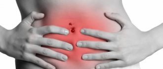
Stool upset, abdominal pain, flatulence are signs of small intestinal disease
Examination of the small intestine is simply necessary to identify polyps, tumors and ulcers. In addition, various types of diagnostics make it possible to identify pathologies of varying complexity at the initial stage of their development. Thanks to the use of various techniques, it is possible to diagnose the problem area, assess the complexity of the disease and determine the course of the operation.
In fact, the small intestine plays an important role in the digestive system. It is in it that the final processes of digestion of basic foodstuffs into relatively simple substances and their subsequent absorption are carried out. Subsequently, the cells of the human body are built from such material.
It is in the small intestine that the absorption of vitamins and minerals necessary for the normal functioning of the body occurs.
Experts identify various pathologies of the small intestine, and each of them has a fairly uniform manifestation. It is for this reason that all digestive problems are combined under the name malabsorption syndrome. Regardless of the cause of the pathology, the development of the following symptoms is observed:
- problems with stool
- rumbling in the stomach
- painful sensations
- flatulence
- intestinal dyspepsia
You can learn more about capsule endoscopy from the video:
Mostly with various disorders of the small intestine, patients complain of stool disorder, which contains remnants of undigested food. The location of pain is usually the navel or pancreas, as well as the right half of the abdomen. Typically, the pain is aching, pulling and bursting in nature, and after the gas has passed, its severity noticeably decreases.
With various ailments of the small intestine, various symptoms appear due to impaired digestion and absorption of basic foods, microelements and vitamins. The patient may rapidly lose weight, lose weight, and cannot gain weight. The result of this is the development of anemia, the appearance of hemorrhages on the body, increased dryness of the skin and disruptions of the menstrual cycle.
Preparation for the procedure
In order for the results of the examination of the small intestine to be correct, it is necessary to properly prepare
To obtain informative research indicators, it is important to comply with certain preparations for any procedure:
- if a capsule is used to diagnose an organ, then this procedure must be performed only on an empty stomach
- if it is necessary to perform some diagnostic tests, laxatives are pre-prescribed to cleanse the intestines
- Before irrigoscopy, you need to empty the intestines of feces using an enema or laxatives, and the procedure itself is carried out on an empty stomach
If endoscopy is necessary, you will have to stop taking medications that contain iron and activated carbon.
Examination using a sigmoidoscope
Used to examine the final sections of the intestine. A sigmoidoscope is a lighting device with a metal tube.
Before insertion, the tube is lubricated with Vaseline for ease of advancement. The procedure allows you to examine the sigma and rectum over a length of 35 cm. Indicated for preventive purposes in elderly patients once a year.
Indications:
- pain in the anus;
- chronic constipation;
- blood, mucus and pus in the stool;
- stool instability;
- foreign body sensation;
- hemorrhoids and colitis.
Contraindications for this procedure are:
- anal fissures;
- narrowing of the intestine;
- peritonitis;
- paraproctitis;
- cardiac pathologies.
The preparation is standard.
Classification of methods
Methods for examining the small intestine make it possible to identify the disease in the early stages and detect complex pathology of any type, even in the absence of pronounced symptoms. Pain and discomfort are minimized.
The most effective methods:
- radiography (using barium);
- endoscopy + capsule examination;
- computed tomography and ultrasound;
- intubation probe.
Any of the above procedures must be preceded by a visit to the doctor. Only a specialist can select the examination method appropriate for a specific patient.
Radiography
X-rays of parts of the small intestine are prescribed to determine enteritis, malabsorption syndrome, peptic ulcers, various tumors, chronic narrowing, and functional malfunctions of the absorption organ. Signs characterizing the development of intestinal pathology are: diarrhea, a sharp decrease in the patient’s weight, black pigmentation of stool, etc.
This procedure uses X-rays to qualitatively evaluate the small intestine. Before the procedure, the patient's intestines are filled with a special pigment substance, which is visible when irradiated with X-rays. It is this that allows you to examine in detail any parts of the suction organ. In most cases, substances such as barium sulfate or barium + air (double contrast) are used for this. The process of moving the contrast agent through the gastrointestinal tract is displayed on the computer monitor. Increased information content is achieved through targeted X-ray images.
A referral for an x-ray is prescribed by an oncologist or therapist. Contraindications to the study apply to pregnant women, nursing mothers and people who suffer from intestinal obstruction
The patient must follow a diet for 2-3 days. Foods that increase gas formation are completely excluded from the diet. The procedure is carried out strictly on an empty stomach. Smokers are advised to abstain from smoking. The doctor must be informed if the patient is taking any medications. It is prohibited to wear various metal objects in the irradiation zone.
Endoscopy
The endoscopic diagnostic method is quite often used when monitoring the small intestine. For a detailed and accurate result, the patient should undergo a complete cleansing using laxatives. In practice, everything happens like this: a specialist inserts a sensor into the rectum, which distributes ultrasound. This is one of the safest methods, which is painless and allows you to detect various diseases (polyposis, obstruction, oncology, etc.). Complications with the heart and lungs are contraindications to the procedure.
Multifunctional capsule
Capsule examination is a minimally invasive diagnosis of the small intestine. This method allows you to examine various parts of the gastrointestinal tract and promptly detect even the initial stages of the disease. All this happens using a special enterocapsule, which is equipped with a universal video camera.
The procedure is carried out if:
- The patient experiences severe pain in the abdominal area.
- Hidden internal bleeding is observed in the body.
- The presence of a tumor and congenital pathologies.
Capsule examination allows you to determine the presence of stomach or intestinal cancer. Like any other diagnosis, a capsule study is carried out after cleansing the gastrointestinal tract of food and on an empty stomach.
Computed tomography and ultrasound
Magnetic resonance imaging is an effective mechanism that allows you to conduct research without using radiography. MRI is considered a safe method. Diagnostics are carried out for the timely detection of pathological changes. Before monitoring, the patient must undergo enema procedures. The patient is also prescribed oral administration of a special contrast agent. The study takes no more than 10 minutes. The device accurately detects the presence of malignant tumors and various types of metastases. Thus, the specialist gets a clear idea of the degree and severity of the ailments.
Ultrasound can also detect diseases in the early stages. When conducting ultrasound diagnostics, the specialist receives information on the computer monitor about the intestines + the trajectory of the organs at a certain moment. In some cases, a universal sensor is inserted into the anus, which clearly records the nature of the tumor, size and localization.
Computer examination methods (MRI, ultrasound) of the intestine are the safest and most informative methods of diagnosis. They do not cause the patient any discomfort or severe pain. Body monitoring is affordable both in price and availability in medical institutions.
Intubation probe
Probing of the small intestine (intubation probe) is the process of penetrating a special tube into the intestinal cavity in order to identify pathological disorders. In this way, samples of intestinal contents can be obtained. The end of the three-channel tubes has universal cartridges, which are made of thin rubber. The third channel has an outlet. The mechanism of action of the device is quite simple. When the equipment is introduced, the cans are filled with air. As a result, the organ shell increases in size. Thus, a section of the small intestine is isolated. The doctor has the practical opportunity to take substances for laboratory examination (biopsy).
An intubation tube is used for intraoperative bowel decompression. In pediatric surgical practice, single-channel and 2-channel devices are most common. To decompress the intestines in infants and newborns, surgeons make a special probe from silicone tubes that soften under the influence of body temperature. Intubation is a rather unpleasant procedure that is performed under anesthesia.
Other techniques
Today, MRI examinations of the intestines are also being carried out. The dye is administered intravenously and through the mouth. The method cannot replace colonoscopy; it is used as an auxiliary one.
The advantage is that it is painless, informative and lacks radiation exposure.
It is based on the magnetic properties of fabrics. When they enter a magnetic field, they give off a resonance, which is detected by the scanner. Diseased areas differ in resonance from healthy ones. Can reveal tumors, inflammation and ulcers. Preparation consists only of taking laxatives and dieting for 2 days.
CT
Computer examination of the intestine is dangerous due to radiation, which is several times higher in level than other radiation methods. Has many contraindications:
- obese patient weighing more than 150 kg;
- renal failure;
- pregnancy and lactation;
- childhood;
- psychosis;
- sewn metal structures into the body.
The technique is highly accurate, and based on its results, a diagnosis can be made without other studies.
Indications:
- inflammation in the gastrointestinal tract;
- polyposis;
- neoplasms;
- bleeding.
Indications for examination of microflora
The study of intestinal microflora is carried out in the following cases:
- stool instability;
- mucus and blood in stool;
- severe flatulence;
- rumbling in the intestines;
- frequent allergies;
- skin with pustules;
- frequent colds.
For the analysis to be correct, medications, enemas, laxatives, and rectal suppositories should be excluded for 3-4 days. Feces are collected in the morning in a volume of 1 tsp. If there is mucus or blood in it, a sample is taken from these places with a spatula. The material must be submitted to the laboratory within three hours.