To verify an oncological diagnosis, it is recommended in all cases to perform a biopsy - intravital removal of material in the form of a piece of tissue from an area in which the presence of a benign or malignant tumor is suspected for the purpose of its subsequent morphological assessment. A biopsy allows not only to confirm or exclude the presence of oncology, but also to recognize the histological type of tumor, which is key to determining further treatment tactics and prognosis.
- Indications for gastric biopsy
- Contraindications to gastric biopsy
- Preparation for the procedure
- Biopsy sampling methods
- Procedure
- How the material is examined
- Possible complications and consequences of a biopsy
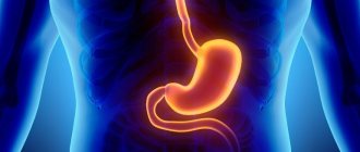
Gastric biopsy is widely used in gastroenterology. It is carried out not only to identify tumors, but also to diagnose other non-tumor diseases. The study is prescribed in combination with esophagogastroduodenoscopy, during which the condition of the mucosa can be assessed and a biopsy specimen taken for histological examination. Previously, gastric biopsy was performed “blindly”, through a gastric tube, but with the introduction of endoscopy, it became possible to accurately take a tissue sample with minimal damage to the mucosa.
Indications for gastric biopsy
Endoscopic examination of the stomach with biopsy sampling is indicated in the following cases:
- Any neoplasms of a tumor nature.
- Sluggish and long-term non-healing ulcers of the mucous membrane.
- Gastritis that is difficult to treat.
- Gastritis with an unclear type of secretion.
- Stomach ulcer.
- Visually detectable changes in the mucous membrane (suspicion of intestinal metaplasia).
- A combination of dyspeptic symptoms and unmotivated weight loss, especially in people with a family history of cancer.
- The presence of polyps of the mucous membrane.
- Previous surgical interventions on the stomach for diagnosed cancer.
Performing a gastric biopsy can serve several purposes:
- Study of the morphological structure of the affected area (differentiation between benign and malignant processes is carried out).
- Determination of inflammatory activity.
- Determination of the type of mucosal epithelial dysplasia.
- Detection of Helicobacter pylori.
- Monitoring the effectiveness of treatment carried out after the diagnosis of stomach cancer.
Indications
Gastric cytology is prescribed to diagnose the following conditions:
The reason for the procedure may also be a tendency to cancer or an inflammatory process. Periodically undergoing cytological diagnostics of the stomach is recommended for people with a predisposition to the listed diseases, with rapid weight loss and poor appetite, in order to monitor and correct the prescribed therapy.
People prone to gastrointestinal diseases need to regularly undergo scheduled examinations to prevent relapses and the development of cancer. After surgery, you need to visit the doctor 2 times a year.
Contraindications to gastric biopsy
Gastric biopsy has a number of contraindications, both absolute and relative. The first group includes:
- Acute somatic pathologies requiring emergency medical care (stroke, myocardial infarction, bronchial asthma attack).
- Cicatricial narrowing of the lumen of the esophagus, making it impossible to advance the endoscope.
- Aortic aneurysm.
Relative contraindications for gastric biopsy include the following conditions:
- Inflammatory process in the nasopharynx.
- Feverish condition.
- Epilepsy.
- Blood clotting disorders, hemorrhagic diathesis.
- Mental illnesses in the acute stage.
- Heart failure and hypertensive crisis.
Preparation for the procedure
No specific preparation is required for the biopsy. It is enough to follow the general rules shown before performing gastroscopy. First, the doctor explains to the patient the purpose and course of the procedure and obtains his consent. In case of emotional lability, before endoscopic examination of the gastric mucosa and taking a biopsy, mild sedatives may be prescribed to calm the patient. In addition, it is necessary to carefully collect anamnesis to identify allergic reactions to drugs used during endoscopy with biopsy (especially anesthetics).
It is recommended to exclude hot and spicy foods and alcohol from the diet a week before the examination, as they can irritate the mucous membranes and distort the picture. You should also reduce your consumption of foods that cause increased gas formation. You should not eat food 6 hours before the procedure, drink 2 hours before. In some cases, it is recommended to rinse the stomach immediately before the biopsy. For example, with pyloric stenosis, the evacuation of food from the stomach into the duodenum may be slowed down.
What does a gastric biopsy show: deciphering the results
What does a regular or HP gastric biopsy show? Why is it being held? How painful and dangerous is this? How to interpret the results? These questions concern every person who has been asked to undergo such a study at least once in his life.
In fact, a biopsy is taking a sample of the mucous membrane, and, if necessary, other tissues of the stomach, for subsequent study of the structure of tissues and cells under a microscope. The resulting samples can be stained with special substances that make it possible to judge the nature of the changes occurring.
A gastric biopsy can show the following changes occurring in this organ:
- atrophy, the nature of changes in the mucous membrane;
- the presence of atypically located cells;
- tumor cell growth;
- type of tumor;
- the type of malignant neoplasm and the degree of its oncogenicity;
- presence of Helicobacter pylori.
Indications for the procedure
Main indications for biopsy:
- suspicion of malignancy;
- precancerous conditions;
- stomach ulcers due to their possible malignancy (cancerous degeneration);
- determination of the type of gastritis;
- H. pylori infection;
- during surgery - in order to control the absence of cancer cells in the remaining part of the organ, determining the type and type of tumor.
Currently, gastric biopsy is performed by performing endoscopy (EGD with biopsy) and by directly taking a tissue sample during surgery.
Surgical method
When performing gastric surgery, the most common method is to excise a sample of pathologically altered tissue with a scalpel.
Endoscopic
Biopsy during gastroscopy can be performed in two ways:
- blind method;
- using visual control during fibrogastroduodenoscopy (FGDS).
With the development of fiber optics, it is the latter method that has become the most popular, because it allows you to take samples from obviously suspicious areas of the mucous membrane and in this way significantly increase the diagnostic value of the study. Using this technique, material is also collected for Hp biopsy (Helicobacter test).
Preparation
General preparation for endoscopic gastric biopsy is practically no different from that during FGDS. The main condition is to come to the procedure room on an empty stomach on the day of the study. For this it is recommended:
- the evening before the sounding, have a light, late dinner;
- in the morning do not have breakfast and do not drink tea, coffee and other drinks;
- Water intake should also be limited and completely stopped at least 2 hours before the procedure.
For several days, it is also prohibited to consume foods and medications that cause irritation of the gastric mucosa, alcohol, hot seasonings and spices. And when prescribing a procedure to identify Helicobacter, antibiotics must also be excluded.
However, in some situations, preparation has its own nuances:
- for pyloric stenosis - gastric lavage before taking a biopsy, since here food can be retained for a day or more;
- children and persons with mental illness - intravenous anesthesia;
- with strong fear of FGDS - injection of atropine + antispasmodic + tranquilizer.
The fibrogastroscope, with which a gastric biopsy is performed, is a flexible probe with a lens and a light source, as well as holes for supplying water and pumping out air.
- Modern devices are also equipped with digital video cameras that display the resulting image on a monitor screen.
- In addition, they have such a structure that they can be used to perform simple endoscopic operations - taking material for examination with biopsy forceps, removing polyps with an excision loop, etc.
The moment of taking a biopsy with an endoscope
An important rule when taking a biopsy of the esophagus or stomach is to take not one, but several tissue samples, preferably from different areas. For example, for gastritis it is necessary to obtain at least 4 samples (2 fragments from the front and 2 fragments from the back wall), and for a tumor or ulcer - 5-8 samples.
- If the procedure is not performed under general anesthesia, then the patient’s oral cavity is irrigated with a 10% lidocaine solution. This is necessary in order to suppress the gag reflex and make further insertion of the probe painless.
- Next, the patient lies on his left side, a special mouthpiece is inserted into his mouth to prevent the jaws from closing, and then the endoscopist gradually inserts a probe through it, examining the esophagus, stomach and duodenum. For better visualization of the mucous membrane, air is simultaneously pumped through the probe, due to which the folds are straightened and the view of the mucous membrane is improved.
- If pathological elements are poorly visible, then in some cases the doctor first irrigates the stomach with a special dye. This may be Lugol's solution, Congo red or methylene blue. Healthy and diseased tissues absorb the dye differently, making tissue sampling sites more visible.
- Next, the biopsy itself is performed. The doctor inserts special forceps through a probe, which bite off small sections of the mucous membrane. In this case, the material is necessarily taken from several places, which increases the likelihood of capturing pathologically altered tissues. The resulting samples are taken out and placed in pre-prepared containers.
- Once the biopsy is completed, the probe is removed and the patient can get up from the couch. It is forbidden to eat food for several hours, but you should abstain from hot foods for a longer period.
The further fate of the obtained samples
- If an urgent answer is needed, then pieces of the biomaterial are frozen and then very thin sections are made with a microtome, which are placed on a microscope slide; if necessary, the resulting material is stained with special dyes and examined under high magnification.
- In standard situations that do not require a quick response, samples are embedded in paraffin, also cut into thin layers with a microtome, stained and studied under a conventional or electron microscope.
- In the case of Hp biopsy 1, the sample is immediately placed in a medium containing urea. H. pylori decomposes it to form ammonia. The presence or absence of a given microorganism is judged by the change in color of the corresponding indicator of the test system. This is a rapid urease test performed during endoscopic examination. The final diagnosis is made when bacteria are detected in tissue sections stained with special dyes.
- In addition, there is a bacteriological method, when the material taken during a biopsy is placed on a nutrient medium that allows the growth of Helicobacter, and the bacterial DNA is also detected in the test sample (PCR diagnostics).
Decoding the results
How long does a biopsy take? If it is urgent, carried out during the operation, then almost immediately, but in standard situations you have to wait 2-3 days for an answer. If samples are sent to another city or country, then the waiting time for a response extends to 1.5-2 weeks.
In the case of a gastric biopsy, deciphering the results obtained is of great importance. In this case, the following parameters are assessed:
- thickness of the mucous membrane;
- epithelium - its character, the degree of its secretion;
- presence of inflammation;
- signs of atrophy, metaplasia, dysplasia;
- degree of H. pylori contamination.
When deciphering the histology of the stomach, one should remember that:
- Sometimes the results may be questionable or unreliable if the amount of material was insufficient, and the study needs to be repeated.
- Gastric cytology is especially important for identifying atypical cells.
- It is the doctor who must ultimately interpret the data obtained.
In general, the results of histological examination can be divided into the following groups:
- Malignant tumors. The type of tumor, the type of cancer cells and the nature of their differentiation (for example, high-, low-differentiated) are determined.
- Benign tumors. The type of tumor and cell type are indicated.
- Gastritis. Its type and the nature of changes in the mucous membrane are described.
- Norm. The stomach tissues are not changed.
Hp biopsy results:
- (-) – negative result, normal;
- (+) – weak contamination, up to 20 H. pylori bacteria in the microscope field;
- (++) – average, moderate contamination, 20-40 bacteria in the field of view;
- (+++) – high contamination, more than 40 H. pylori in the field of view.
Contraindications
A biopsy is completely contraindicated in the following situations:
- acute stroke, heart attack;
- attack of bronchial asthma;
- narrowing of the esophagus that is impassable to the probe (stenosis).
Relative contraindications to endoscopic manipulations:
- fever;
- epilepsy;
- hypertensive crisis;
- hemorrhagic diathesis;
- acute pharyngitis, tonsillitis or exacerbation of chronic;
- heart failure.
Possible complications
As a rule, biopsies performed during EGD rarely cause serious complications. Typically, patients may feel slight discomfort in the stomach area in the first hours after undergoing the test. In addition, minor bleeding from the resulting lesions in the area where samples are taken is possible, and this will go away on its own.
However, if the following signs appear, you should definitely see a doctor or call an ambulance:
- brown vomit, similar in color to coffee grounds;
- nausea, pain in the stomach;
- stomach ache;
- increased temperature, fever;
- severe weakness, increased fatigue;
- sharp deterioration in general condition;
- inflammation of the mucous membranes of the oral cavity, nasopharynx;
- difficulty breathing, chest pain.
These symptoms may signal the following rare but serious complications:
- severe bleeding that does not go away on its own;
- infection;
- septic shock;
- aspiration pneumonia;
- damage to the integrity of the stomach, duodenum, esophagus.
(4 4,50 of 5) Loading...
Source: //GastroMedic.ru/diagnostika/chto-pokazyvaet-biopsiya-zheludka.html
Biopsy sampling methods
A tissue sample from the gastric mucosa can be obtained by two methods. The first method is surgical. It is carried out directly during an operation on an organ, when the question arises about the volume of tissue to be removed. A small section of tissue is removed with a scalpel and sent urgently to the laboratory. The doctor examines the drug under a microscope and makes a conclusion after 10 - 15 minutes. During this time, the operating team does not perform any actions. After deciphering the biopsy, the operation resumes and the necessary adjustments are made to the extent of the surgical intervention.
The more common method is endoscopic. For this purpose, a flexible probe is used - a fiberscope, at one end of which there is a camera lens, an LED flashlight, holes for air and water supply, and a mount for attachments. At the other end of the tube there is a handle with a control unit and an eyepiece. Nowadays, targeted biopsy is mainly used because it minimizes the risks of the procedure.
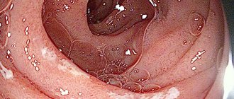
Stomach polyp
Gastric polyp (synonyms: adenoma, fibroadenoma, adenomatous polyp) is considered a benign tumor arising from the integumentary pitted epithelium. In patients with gastric polyposis, the mucous membrane remains intact very rarely. Usually, changes characteristic of atrophic-hyperplastic gastritis or “restructuring” gastritis are observed.
Cytological preparations reveal a large number of layers and accumulations of integumentary pitted epithelium. With adenomatous polyps, glandular structures are also found.
Compared to cytograms for gastritis, in these diseases there is a slight predominance of young, immature cells with enlarged light nuclei and a delicate mesh-like chromatin pattern in them (See Fig.). There are single mitotic figures.
Glandular epithelial cells and lymphoid elements are usually found in small quantities. It is not possible to judge the structure of the glands from cytological preparations. In cases of granulation polyps with abundant inflammatory infiltrates in their stroma, significant numbers of neutrophilic leukocytes can be included in cytological preparations.
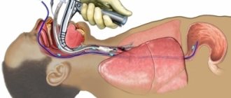
Procedure
A gastric biopsy can be performed on an outpatient basis, under local anesthesia: the pharynx and pharynx are irrigated with a 10% lidocaine solution to suppress the gag reflex. The subject should be in a lying position on his left side. The endoscope is inserted with gentle movements, and all areas of the mucous membrane along the esophagus and stomach are examined one by one. To improve visualization, air is injected, which straightens the folds of the hollow organ.
A piece of tissue is pinched off from the area of mucosa being examined with a special biopsy instrument (biopsy forceps) inserted into the instrument port of the fiberscope. To increase the accuracy of histological examination, material is collected from several points of the mucosa (at least 4), and the biopsy sample is taken from the border between the pathologically changed and normal areas.
An additional technique of chromatogastroscopy is also used, which simplifies the determination of the boundaries between different areas of the surface under study. A dye is sprayed over the gastric mucosa, which is unevenly absorbed (depending on the functional and organic state of the mucosa). For staining you can use Lugol's solution, methylene blue, Congo red. The area that is stained more intensely is most often pathologically changed, and the biopsy is performed on the periphery of this area (if there is an ulcer or tumor, then in the center too). The duration of the procedure can vary from 10 to 40 minutes and depends on the scope of the study.
After a gastric biopsy, it is advisable not to eat for 2 hours. After this period, you should avoid eating hot foods and drinks for some time. As a rule, there is no pain either during or after the procedure, but there may be slight discomfort in the epigastric region. In rare cases, there may be slight bleeding that stops on its own.
Cytological examination of biopsy material for Helicobacter pylori
A cytological test for Helicobacter pylori
is a laboratory test of biopsy material for the content of Helicobacter pylori bacteria to identify
gastroenterological diseases
.
The material is extracted from the mucous membrane of the digestive organs using fibrogastroduodenoscopy
(
FGDS
).
| Deadlines | 5-7 days |
| Synonyms (rus) | Biopsy for H.Pylori, endoscopy for H.Pylori, smear for H.Pylori, HP test, urease test |
| Synonyms (eng) | Biopsy for H. Pylori, endoscopy for H. Pylori, smear mark on H. Pylori, HP-test, urease test |
| Preparing for the study | For an accurate picture of the laboratory test, the biopsy is performed strictly on an empty stomach. |
| A study of biopsy material is performed after stopping antibiotics, drugs that thin the blood or increase clotting, before using antibacterial drugs or one and a half weeks after stopping the drug | |
| 10 hours before the test you should refrain from eating and limit drinking | |
| Type of biomaterial and methods of taking it | Tissue of the stomach and esophagus obtained by biopsy (FGDS). |
What is a cytological test for Helicobacter pylori?
Helicobacter pylori
(Helicobacter Pylori) is a flagellated bacterium that lives on the mucous membrane of the gastrointestinal tract. This bacterium is highly active and today more than 60% of the population of industrialized countries and up to 90% of developing countries are infected with it.
Helicobacter infection
is the main cause of
stomach ulcers
.
The bacterium is transmitted by contact. Today it has been revealed that men suffer from ulcers 4 times more often than women, and people with the first blood group are an order of magnitude more likely than everyone else. Infection with the bacterium is a cofactor in the occurrence of gastric adenocarcinoma
(
stomach cancer
).
Cytology is the most accurate method for determining bacteria in the stomach. With proper preparation for the examination, a false result is practically excluded. The analysis is intended for patients with suspected diseases of the digestive system
caused by Helicobacter bacteria: gastritis, stomach ulcers, cancer.
The material used is a smear-imprint obtained during fibrogastroduodenoscopy
. Biopsy material is removed from suspicious areas of the gastric mucosa, excluding grossly damaged areas, since Helicobatera does not live in ulcers and erosions. The material is applied to glass and delivered to the laboratory for research.
Interpretation of the results of cytology of gastric biopsy
The results of a mucosal biopsy can have two qualitative meanings:
- negative result (this is normal, no bacteria are detected);
- positive result (if at least 1 bacteria is detected).
In quantitative terms, a positive result can take the following values:
- low contamination (+) – there are up to 20 Helicobacter pylori in the field of view;
- moderate contamination (++) – the number of bacteria in the smear reaches 40;
- high contamination (+++) – more than 40 bacteria were found in the smear.
If a patient is examined for the first time and receives a positive result, then all his close relatives are recommended to undergo the examination and the value of the titres does not matter.
If low values are obtained, it is recommended to confirm the analysis by other methods: stool antigen analysis, helic tests, blood tests. If the results are confirmed, it is worth undergoing serious treatment, since Helicobacter pylori is a rather dangerous bacterium that can cause severe damage to the internal digestive organs.
Upon cytological examination
of the material obtained from the stomach using gastrobiopsy, the cellular elements of the gastric mucosa are clearly identified in the preparations: the integumentary pitted epithelium, the main and parietal cells of the glands.
Ratio in cytological preparations
cellular elements of the gastric mucosa, varying degrees of their differentiation, the appearance of signs of degeneration and atypia, as well as the appearance of leukocytes, reticular and histioid elements make it possible to identify the types of gastrocytograms. which are most characteristic of one or another stomach disease.
Gastrocytograms
patients with chronic gastritis are characterized by the presence of cells of the integumentary pitted epithelium, which are located predominantly in small layers and clusters. The cells have a high and low cylindrical shape, a basal or centrally located nucleus. wide, slightly basophilic cytoplasm. The cell nuclei are distinguished by some polymorphism; there are all transitions from immature, relatively large, light chromatin with a delicate mesh pattern to mature small sized, intensely colored nuclei.
Chief and parietal cells of glands
found in varying quantities, located scatteredly, in small rounded groups or even rows, in which the correct alternation of main and parietal cells is noted. In the main cells, coarse granules colored blue-violet are clearly visible, filling the entire cytoplasm. The parietal cells are distinguished by a wide, light cytoplasm, painted in a soft pink color. The preparations contain lymphoid elements and polymorphonuclear neutrophil leukocytes.
How the material is examined
The piece of tissue taken during the biopsy is placed in an airtight container with a preservative. The container is signed (full name of the person being examined and the date of the procedure) and sent to the laboratory.
In the histological laboratory, the tissue is fixed in formalin and embedded in paraffin; if an urgent examination is performed, the tissue is frozen. Then the laboratory assistant uses a microtome to make thin sections of the tissue being examined. The specimen is then placed on a glass slide, stained, dried, and examined under a microscope.
After studying the drug, the pathologist issues a conclusion in which he indicates:
- thickness of the gastric mucosa;
- the nature of the epithelial tissue;
- the presence of dysplasia or metaplasia of the epithelium;
- degree and nature of inflammation;
- the presence of atypical cells, their maturity and location;
- presence of H. pylori, degree of contamination.
A possible additional analysis may be the polymerase chain reaction of a biopsy specimen, which gives the most accurate result in terms of diagnosing Helicobacter pylori.
Morphologically, the presence of the following signs speaks in favor of stomach cancer: abnormal cell shape (polymorphism not typical for this area), cell size (usually tumor cells are larger), size and fragmentation of nuclei, multinucleation, pathological inclusions in the cytoplasm.
The results of a gastric biopsy, if it is not urgent, are given to the patient and the attending physician 10 to 14 days after the procedure. An urgent study is performed on the same day, but its accuracy is somewhat inferior to a planned study.
Gastric biopsy results: methods and interpretation
The average turnaround time for gastric biopsy results is from 5 to 14 days, depending on the workload of the pathology laboratory. At the end of the histological examination, the attending physician (sometimes the patient himself) receives a conclusion from a pathologist with a transcript on the basis of which treatment tactics are planned.
This article presents a transcript of the results of a gastric biopsy with a detailed description of each term that can be found in the conclusion.
Biopsy techniques
In most cases, a small sample of the gastric mucosa obtained during gastroscopy is sent for pathohistological examination. Already in the laboratory, histological preparations are made from this fragment with a diameter of 3-5 mm, which are stained using various methods.
The benefits of gastroscopy with gastric biopsy are on the patient's side. This procedure is low-traumatic, minimally invasive and in some cases is not accompanied by any discomfort. However, from the point of view of a pathologist, such small gastric biopsies have a number of disadvantages:
- Often the wrong fragment is sent for examination: during gastroscopy, the stomach continues to contract and the endoscopist needs to try to “catch” the area of the mucous membrane that interests him;
- It is quite difficult to detach a polyp or a section of mucous membrane without damaging the tissue;
- The vast majority of biopsy samples are sent crumpled and even crushed, which increases the risk of diagnostic error;
- Due to the small size of the biomaterial, it is impossible to accurately determine the histological type of the tumor and the extent of its spread if cancer cells are found in the sample.
Interpretation of biopsy results
The conclusion usually provides a description of the histological picture (cellular and tissue structure of the biopsy) and the pathomorphological diagnosis. Despite the existence of a clear method for deciphering the results of a gastric biopsy, many doctors give an insufficiently complete description. The table presents the main terms that can be found in the results and their characteristics:
| Term | Description |
| Helicobacter pylori (Helicobacter pylori, HP ) | Bacteria, the leading cause of chronic gastritis, peptic ulcers and stomach cancer. In many laboratories, it is assessed using a system of crosses: 0 – no CP, from 1 to 3 crosses – the presence of bacteria in one quantity or another. |
| Adenocarcinoma | Name of stomach cancer. |
| Adenoma | Benign neoplasm of epithelial cells of the mucous membrane. In structure it can be tubular (in the form of tubes), papillary (in the form of finger-like processes) and mixed. Adenocarcinoma often develops against the background of adenoma. |
| Activity | A conditional indicator reflecting the degree of inflammation of the mucous membrane. Assessed by the number of neutrophils, leukocytes, degree of metaplasia and atrophy. Divided into 4 degrees: 0 – no inflammatory activity, 3 – severe gastritis. |
| Antrum | Section of the stomach, represented by convoluted, closely located glands. |
| Atrophy | Thinning of the mucous membrane, disappearance of folding. It is graded using a cross system from 0 (no atrophy) to +++ (complete atrophy). |
| Goblet cells | Cells that produce mucus. Normally they are not present in the stomach. A common component of intestinal metaplasia. |
| Hyperplasiogenic polyp | Benign growth of the mucous membrane due to pronounced regeneration (recovery) against the background of inflammation. |
| Hyperplastic polyp | In general, an analogue of a hyperplasiogenic polyp. |
| Purulent-necrotic masses | Accumulations of neutrophils and dead cells. Often found at the bottom of an acute ulcer. |
| Cardiac department | A section of the organ that is similar in structure to the antrum, but the glands are more loosely located. |
| Cribrotic structures | Sign of cancer: glands with common walls (normally, each gland has its own walls). |
| Lymphoid infiltration | The presence of lymphocytes, plasma cells and macrophages in the biopsy specimen. A sign of a chronic inflammatory process (gastritis). Evaluated according to the cross system. |
| Lymphoplasmacytic infiltration | The same. |
| Malingization | Malignancy. This means that in some part of a benign tumor there is a cluster of cancer cells. |
| Metaplasia | Replacement of normal gastric epithelium with intestinal epithelium. There are small intestinal and colonic metaplasia, and the latter poses a great danger in terms of cancer development. |
| Mononuclear cell infiltration | The same as lymphoid. |
| Neutrophil infiltration | Accumulation of segmented neutrophils. A sign of acute gastritis and stomach ulcers. Evaluated using a cross system from 0 to +++. |
| Paneth cells | Cells characteristic of the mucous membrane of the small intestine. Occurs with metaplasia of the small intestinal type. |
| Signet ring cells | Cells with a visible “void” in the center, which is why they look like rings under a microscope. Sign of stomach cancer. |
| Pyloric glands | Glands that are located in the final sections of the stomach. |
| Skirr | Cancer with severe fibrosis (overgrowth of connective tissue). |
| Stroma | The connective tissue base in which the glands are located. |
| Body of stomach | Middle section with straight glands and clearly visible parietal cells (which produce hydrochloric acid). |
| Fibrous tissue | Dense connective tissue. Develops against the background of chronic inflammation, occurs in the bottom and walls of ulcers, as well as in cancer. |
| Fibroplastic reaction | Overgrowth of connective tissue. Often found in descriptions of cancer. |
| Foveal hyperplasia | An increase in the number of normal cells located in the mucosal pits. |
Following actions
If the results of a gastric biopsy contain terms such as “atypical cells”, “cribrous structures”, “signet ring cells”, “carcinoma”, “cancer” and others, then you should not panic.
To clarify the diagnosis, it is recommended to do a repeat gastroscopy with a biopsy (preferably in another clinic and with another doctor) and if oncological pathology is detected again, then only in this case should you contact an oncologist.
In the most modern Israeli clinic Top Ichilov , thanks to the use of the latest techniques and drugs, more than 90% of the clinic’s patients suffering from stomach cancer recover completely or achieve significant improvement and long-term remission.
Treatment of stomach cancer in Israel is carried out by world-class specialists who practice the latest methods of fighting cancer: chemotherapy, immunotherapy and biotherapy using the latest drugs that are selected individually for each patient in accordance with the results of advanced molecular genetic tests, a unique HIPEC chemotherapy technique that allows achieve better results and avoid side effects, the Da Vinci robotic surgical unit, with the help of which microsurgical operations of increased complexity are performed, and many others. You can find out more about treatment in Israel by filling out a request in the form below.
If a study was carried out on H. pylori and a biopsy confirmed the presence of these bacteria, complex eradication therapy using 3-4 drugs is prescribed. After 10-14 days, FGS must be repeated to evaluate the effectiveness of treatment.
Source: //ProBiopsii.ru/rasshifrovka-rezultatov-biopsii-zheludka/
Possible complications and consequences of a biopsy
With proper preparation and a biopsy performed by a qualified doctor using modern equipment, the risk of complications is significantly reduced. However, in practice the following situations may rarely occur:
- acute bleeding from the area where the material was taken;
- perforation of the gastric mucosa. It occurs extremely rarely, usually when a “blind” biopsy is performed by an inexperienced specialist);
- the occurrence of allergic reactions to the anesthetic;
- damage to teeth and oral mucosa.
In addition, after the procedure, long-term consequences may occur, which, as a rule, are facilitated by concomitant pathology, as well as some anatomical features:
- bleeding from a vessel damaged during biopsy;
- aspiration pneumonia due to violation of the technique of inserting and removing the fiber gastroscope or vomit entering the respiratory tract;
- inflammatory diseases of the upper digestive system.
A gastric biopsy is in most cases a painless and low-traumatic procedure, which, despite its relative simplicity, can provide a lot of important information about the nature and course of the pathological process.
Book a consultation 24 hours a day
+7+7+78