Even in the recent past, doctors could rely solely on symptoms and indirect signs of the disease to make a diagnosis. It was possible to see the pathology with one’s own eyes only during an autopsy, after the death of the patient. Fluoroscopic research methods have become undisputed “helpers” in diagnosis and treatment. However, they could not provide complete information about the condition of the hollow organs (stomach and intestines), since most diseases of the gastrointestinal tract cause, first of all, internal damage to the mucous membrane, which cannot be seen on an x-ray, and only in extreme cases cause deformation of the external or internal contour of an organ (for example, during the formation of a tumor).
Today it is possible not only to examine in detail the internal structure of the stomach and duodenum, but also to do this in the least traumatic way for the patient. An example of such a diagnostic procedure is an alternative to classical gastroscopy – gastroscopy through the nose.
What it is?
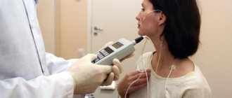
Transnasal gastroscopy is an FGDS method in which an endoscope is inserted through the nasal passage instead of the oral cavity . The probe then passes through the back wall of the nasopharynx and oropharynx, the larynx and enters the esophagus. At its end there is a small video camera with lighting, the image from which is shown on the screen. This allows the doctor to control the progress of the device, as well as examine the mucous membrane and contents of the upper part of the digestive tract. The endoscope also necessarily has a sensor that measures the acidity of the environment.
If necessary, with transnasal gastroscopy, as with conventional FGDS, of the altered area of the mucous membrane can be performed This sample is then sent to the laboratory for cytological examination.
Help The diameter of the endoscope, which is used for transnasal insertion,
is 7-7.5 mm , which is significantly smaller than that of a conventional probe (9-10.5 mm). That is why the procedure is much easier to tolerate.
How is the procedure performed?
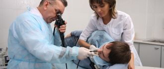
Nasal gastroscopy is performed to examine the gastric mucosa to make the correct diagnosis. During the study, a special apparatus is used, which is equipped with a camera and a flashlight. The image is displayed on the screen, and the specialist can examine in detail the condition of the internal organ.
During the examination, the following pathological processes are revealed:
- ulcers;
- bleeding;
- inflammation;
- bacterial activity.
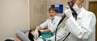
The device is inserted through the nasal sinuses, enters the nasopharynx and is inserted into the stomach. Using FGS, you can examine the stomach and duodenum. To carry out the procedure in this way, a special device is used, different from the classic sensor for FGDS. Modern devices are quite thin, so the patient will feel absolutely no discomfort, and the likelihood of complications is reduced to zero.
The procedure has some features:
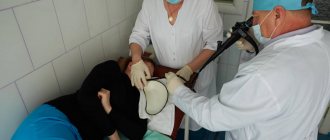
- To perform gastroscopy, the patient must lie on his side. The sensor is inserted into the nostril, the second one remains free, so the patient breathes normally.
- In order to minimize discomfort, the specialist treats the mucous membrane with a local anesthetic.
- To ensure that the tube enters as comfortably as possible, a special gel is used to insert it.
- The event must be performed on an empty stomach.
- If you have a runny nose, the nasal cavity is first cleared of mucus.
- It is important that the patient can freely communicate with the doctor during the procedure, reporting his feelings.
- Discomfort can only occur when the doctor turns the sensor in one direction or another.
Modern research methods allow the procedure to be carried out painlessly and quite quickly. In this case, the specialist receives a complete picture of the state of the digestive organs.
Indications for transnasal EGD
FGDS is one of the most commonly used diagnostic techniques in gastroenterology. It is prescribed if the patient has the following symptoms:
- aching or sharp cutting pain in the upper abdomen, which intensifies after breaking the diet, drinking alcoholic beverages, or pressing with fingers;
- a feeling of heaviness in the stomach and its rapid filling during meals;
- sudden loss of appetite;
- nausea and vomiting with blood , bile or more mucus;
- tendency to constipation or diarrhea;
- sensation in the chest or frequent heartburn;
- weight loss without changes in diet or physical activity;
- belching sour;
- the appearance of a white coating on the tongue or mucous membranes of the mouth;
- a sharp change in the patient’s taste preferences (the appearance of an aversion to meat).
In addition, for a number of conditions, transnasal gastroscopy is preferable or is the only possible choice. These include:
- acute inflammatory processes of the oral cavity;
- impaired swallowing (consequence of a stroke or tumor in the central nervous system);
- aneurysm (expansion or protrusion of the wall) of the thoracic aorta;
- narrowing of the esophagus;
- severe nausea during routine FGDS;
- increased anxiety and fear of the patient before the procedure.
Description of the technique
The need to develop a new direction in endoscopy of the upper gastrointestinal tract was dictated primarily by the stress factor in patients. Many of them, during a classic transoral examination (EGD), experience a strong fear of suffocation (asphyxia) or getting contents into the respiratory tract (aspiration).
Great importance is also given to negative experiences during previous episodes of endoscopy, which became the reason for the use of sedative (calming) drugs. They have side effects on the respiratory and cardiovascular systems, so for the majority of patients they are limited.
Transnasal gastroscopy differs in that an instrument with a camera is inserted through the nasal passages, meaning the person does not need to swallow the probe.
In this case, classic local anesthesia of the pharynx is used using a 10% solution of Lidocaine (usually in the form of an optically transparent gel). 10 minutes before this, the lower nasal passage that will be used is irrigated with a spray with mucolytics and vasoconstrictors.
Indications
This technique still does not have critical differences, therefore fibrogastroduodenoscopy (FGDS) through the nose is indicated in the same cases as with transoral insertion of the instrument. These include the following:
- Presence of typical gastrointestinal complaints (heartburn, nausea, pain in the upper abdomen, vomiting);
- Gastritis;
- Varicose veins of the esophagus;
- Peptic ulcer of the stomach, duodenum;
- Polyposis;
- Acute abdominal pain syndrome;
- Ineffectiveness of the therapy received;
- Monitoring the dynamics of the disease;
- The impossibility of performing transoral FGDS is a strong gag reflex, fear of asphyxia, severe heartburn, extensive damage to the oropharynx, etc.;
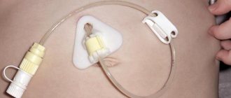
- In some cases, patients require placement of a nasogastric tube (pre- and postoperative period, intensive care).
An endoscopic examination allows you to visually assess the condition of the gastrointestinal tract, as well as carry out other manipulations - biopsy, polypectomy, stopping bleeding.
Contraindications
The use of nasal gastroscopy is not recommended in the following situations:
- Clinically significant obstruction of the nasal passages;
- Deviation of the nasal septum, injuries to the skull bones;
- Acute phase of respiratory tract diseases (rhino-, adeno-, enterovirus infections, influenza);
- Unstable condition of the patient (psychoneurological pathology);
- Narrowing of the esophagus - strictures, stenosis;
- General serious condition of the patient;
- Massive internal bleeding.
Only the attending physician is able to correctly assess the patient’s condition. In some cases, all indications/contraindications may become relative if the diagnostic benefit outweighs the health risk.
Preparation for gastric endoscopy
Preparatory measures before transnasal endoscopy are almost identical to those before conventional FGDS. The procedure must be carried out on an empty stomach, so the patient must have his last meal 12 hours before the expected start. An hour before FGDS, fluid intake is also limited. You can read in detail about preparing for the procedure here.
Please note: Before the examination,
the patient often needs to be examined by an otolaryngologist (especially if there is a history of rhinitis, tumors or trauma to the nasal cavity). This makes it possible to identify patients with a contraindication to the procedure.
What is the best way to insert the probe?
Transnasal endoscopy is increasingly used as an alternative to the traditional FGDS technique. Their main advantages and disadvantages are summarized in the following table:
| Advantages | Flaws | |
| Transnasal gastroscopy | Less severe discomfort for the patient; · voice contact with the patient is maintained; · more free breathing; · Possibility of carrying out in case of narrowing of the esophagus or difficulty swallowing | · less accessibility (the necessary probe is not available in all clinics); Possibility of injury to the nasal mucosa |
| FGDS with insertion of a probe through the mouth | · greater availability; · opportunities for minimally invasive interventions (for example, stopping bleeding) after diagnosis | · more pronounced dyspeptic symptoms (abdominal discomfort, nausea); · lack of voice contact with the patient; · higher risk of injury to the throat mucosa ; Possible breathing difficulties (psycho-emotional in nature) |
Advantages and disadvantages
Gastroscopy through the nose is currently the most popular method for examining the upper gastrointestinal tract due to the following advantages over the transoral method:
- There is no contact of the gastroscope with sensitive areas, so there is no gag reflex and no urge to swallow the instrument;
- During the procedure, the person can talk to the doctor, since there is no need to use a special mouthpiece. This increases the level of information content of the survey;
- The patient does not experience pain or discomfort, his emotional state remains satisfactory, so sedatives are not used;
- Typically, transnasal gastroscopy takes less time than with the traditional technique;
- At the end of the procedure, the person immediately feels normal and can drive or go to work;
- No need to diet. After the examination, you can immediately eat;
- Treatment costs are reduced, since fewer medications are used and there is no need to monitor the patient’s condition;
- The latest ultra-thin video endoscopes are used, which are highly maneuverable and easy to insert. Such equipment is usually equipped with better resolution cameras and other parts that allow minor interventions or biopsies to be performed.
- The small diameter of endoscopes determines the use of nasal gastroscopy in patients with stenosis and children.
Despite the positive aspects, the method has the following disadvantages:
- There is a risk of damage to the mucous membrane of the nasopharynx, which could potentially lead to bleeding;
- During the study, other medications are taken - “defoamers”;
- Given the small diameter of the gastroscope, the scope of minimally invasive interventions is limited.
Contraindications
Despite all the advantages of transnasal gastroscopy, there are a number of conditions when its implementation is strictly prohibited. These include:
- tumors of the nasal cavity or nasopharynx;
- consequences of traumatic injury to the nose (for example, deviated nasal septum);
- infectious processes that are accompanied by swelling of the nasal mucosa ;
- frequent nosebleeds;
- traumatic brain injury;
- instability of the patient's general condition;
- condition after myocardial infarction, or unstable angina;
- blood clotting disorder (congenital or acquired);
- mental disorders with inadequate perception of medical procedures;
- bronchial asthma in the acute stage;
- disturbance of external respiration;
- acute phase of ischemic or hemorrhagic stroke;
- cardiac arrhythmias, which can threaten the patient’s life, without proper drug therapy or installation of an artificial pacemaker.
Possible consequences
Complications may also develop during transnasal gastroscopy. The most common of them are collected in the following table:
| Complication | First aid at home |
| Nausea | Refrain from eating for 2-3 hours after the procedure, take a sitting position, open the window, breathe deeply and slowly |
| Discomfort in the abdomen | Follow a diet, drug therapy (antacids, proton pump inhibitors) |
| Feeling of dryness in the nose | Water inhalations, use of sea water-based spray |
| Nasal congestion | Use vasoconstrictor drops (based on xylometazoline, oxymetazoline) |
| Nose bleed | Bend over the washbasin so that the blood can flow out, and place a handkerchief soaked in cold water or ice on top of your nose. If this does not help stop the bleeding, use a narrow piece of bandage to tamponade the nasal passage (close it completely and tightly) |
| Sharp cutting pain in the abdomen | Lie on your back, avoid excessive physical activity, call an ambulance or a doctor |
| Heart rhythm disturbances (the occurrence of atrial fibrillation or flutter) | Avoid excessive physical activity, take antiarrhythmic drugs (amiodarone, propafenone, flecainide), seek medical help |
| Feeling of irritation in the throat | Use tablets with local anesthetics and/or anti-inflammatory components, rinse with herbs (calendula, chamomile) |
| The appearance of a cough and a sharp increase in body temperature (signs of stomach contents entering the bronchi and lungs) | Seek medical help (you must undergo a chest x-ray and a course of antibiotic therapy) |
Alternative Methods
If transnasal gastroscopy is impossible, or its information content is insufficient, other methods of diagnosing the upper part of the digestive system are prescribed:
- Ultrasonography. Allows you to detect congenital and acquired anomalies, large tumors of the stomach and duodenum.
Computed tomography is a radiological diagnostic method (sometimes using contrast) with high information content. Often used to diagnose oncological pathologies of the stomach and their metastases.- Magnetic resonance imaging is a non-invasive diagnostic method that is based on the principle of nuclear magnetic resonance. Allows highly informative visualization of the abdominal organs.
- Video capsule endoscopy . The patient swallows a small capsule-shaped chamber that sequentially passes through all parts of the digestive system.
- X-ray or fluoroscopy of the stomach (using contrast). Makes it possible to check the contours and integrity of the organ.
The essence of the method
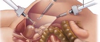
Gastroscopy, today, is the most informative diagnostic method, allowing not only to examine the mucous membrane of the upper gastrointestinal tract (GIT), but also to perform minor surgical procedures, in particular:
- removal of small benign neoplasms (polyps, papillomas);
- taking samples of the mucous membrane for histological analysis if malignant tissue degeneration is suspected;
- the use of high-frequency currents to cauterize injuries in order to prevent bleeding;
- suturing cracks and tears.
All this became possible thanks to the development of a fiber-optic system, which is the main structural element of the gastroscope. It is with the help of a bundle of the finest fibers that the image from the distal end of the gastroscope is transmitted to the monitor, allowing a variety of manipulations to be performed under visual control. As a rule, a gastroscope has a standard length of about 130 cm and a thickness ranging from 0.5–1.3 cm.
A significant design difference between gastroscopes is the location of the optical system; it comes in the following types:
- frontal;
- lateral;
- oblique.
The lateral position is used to examine the blind areas of the duodenum during esophagogastroduodenoscopy. However, due to the good flexibility and controllability of the distal end, in most cases gastroscopes with frontally located optics are used in practice.
Fiber gastroscope with frontal optics
The term gastroscopy includes two types of diagnostics:
- fibrogastroscopy (FGS), during which the esophagus and stomach are examined;
- esophagogastroduodenoscopy (EGDS) or fibroesophagogastroduodenoscopy (FEGDS), including the duodenum in the list of objects examined.
Performing gastroscopy using the classical method involves introducing an endoscopic system through the patient’s oropharynx into the esophagus. It is this part of the procedure that usually causes many difficulties that complicate the doctor’s work and frighten the patient. Transnasal gastroscopy allows you to avoid a number of difficulties associated with side effects that arise during the process of swallowing the probe.
Important! A limitation to transnasal gastroscopy may be a too narrow nasal passage or a deviated nasal septum, which does not allow insertion of a probe into the opening.