Anatomy of the human gastrointestinal tract
The human gastrointestinal tract (its anatomy is a collection of organs designed to carry out the digestion process) has an average length of 8 to 10 m.
The gastrointestinal tract performs the following functions:
- Motor. Consists of moving food through the organs of the tract with the help of muscles.
- Secretory. It represents the production of juices by glandular cells.
- Suction function. It is the process of penetration of nutrients and fluids into the bloodstream.
- Excretory (excretory).
- Endocrine (formation of various hormones - gastrin, secretin).
Diseases of the gastrointestinal tract
Many diseases of the gastrointestinal tract are caused by disturbances in the functioning of the immune system, which cannot cope with the number of harmful factors that it encounters daily. And if a person has a genetic predisposition, coupled with poor nutrition, tobacco and alcohol abuse, then gastrointestinal diseases will not take long to appear. Let's look at the most common diseases of the digestive system.
Stomatitis is a disease that affects the mucous membrane of the mouth. It can cause very unpleasant sensations. As a result, the ability to chew food well is reduced, which ultimately has a detrimental effect on the functioning of the stomach. The cause of stomatitis is weak immunity.
Esophagitis occurs when the lining of the esophagus becomes inflamed. This can be caused by drinking alcohol, too rough, poorly chewed food, or burns. Diseases of the gastrointestinal tract, such as esophagitis, cause quite severe pain and discomfort. There may be a burning sensation, vomiting, sometimes even mixed with blood.
A huge number of people suffer from heartburn. This condition is associated with increased acidity of gastric juice. When part of it rises into the esophagus, a burning sensation occurs.
Chronic gastritis is the most common gastrointestinal disease. Previously, it was believed that gastritis is a disease of students and people with a frantic pace of life who eat irregularly and incorrectly. Today it is absolutely known that the vast majority of gastritis is caused by the bacterium Helicobacter pylori. Helicobacter pylori infection is one of the most common in the world and speaks volumes about how poor the state of the immune system is in most people. Chronic gastritis is an inflammation of the gastric mucosa. In fact, it is a disease that can have very serious consequences. First, the absorption of various beneficial substances, for example, vitamin B12, is impaired. A deficiency of this vitamin leads to the development of anemia. If gastritis is not treated, its atrophic form may develop, which is considered a precancerous condition.
Chronic duodenitis and chronic colitis are inflammations of the mucous membranes of the duodenum and large intestine, respectively.
These are not all diseases of the gastrointestinal tract. There are many more of them, some of them are very dangerous, such as peptic ulcer disease or pancreatitis. Of course, the ideal option is prevention, which will help prevent gastrointestinal diseases. But what to do if the diseases have already been diagnosed?
Structure and functions of the gastrointestinal tract
Different sections of the gastrointestinal tract have different structures and perform strictly defined functions.
Oral cavity
In front, the oral cavity is covered by the lips. On the sides it borders with the cheeks, and is separated from above by the hard palate. At the back is the pharynx, which leads to the pharynx.
The functions of the oral cavity are food grinding and enzymatic function.
Pharynx
This is an unpaired organ that looks like a funnel. The pharynx is located between the oral cavity and the esophagus. In front of the pharynx is the larynx, nasal cavity and oral cavity. It is attached superiorly to the occipital bone. The top of the pharynx is called the fornix. The pharynx is divided into the nasopharynx, oropharynx and laryngopharynx.
Esophagus
It is a thin muscular tube. In the cervical region it is narrowed, then gradually begins to expand, moving into the abdominal part. The wall of the esophagus is lined with mucous membrane, submucosal layer and muscles.
Stomach
In its structure, it has 2 walls (back and front) and 2 holes. A greater and lesser curvature is formed along the posterior wall of the stomach. The stomach has a cardiac opening through which it communicates with the esophagus. The second exit is the gatekeeper area.
The convex opening of the stomach is represented by the fundus of the stomach. The body of the organ is located between the bottom and the pylorus.
The wall of the stomach is formed by the mucous membrane, which forms folds. On the mucous membrane there are pits in which the glands pass. The stomach is covered with mucous, submucosal, muscular layers and serous membrane.
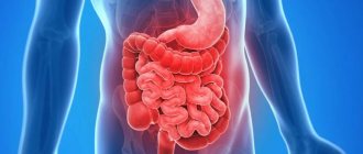
Intestines
The human gastrointestinal tract (anatomy includes the entire intestine) is complex. The small intestine is represented by the cecum, colon, sigmoid and rectum. The small intestine consists of the duodenum, small intestine and jejunum.
Anal hole
The anus is the final anatomical formation of the digestive tract. It is surrounded by sphincters, which are formed from muscle fibers: the internal one, which is formed by smooth muscles, and the external one, consisting of striated muscles.
Auxiliary organs
In addition to the main ones that are directly part of the digestive system, auxiliary organs also play an important role.
Liver
The liver is the largest gland in the human body. It is located in the right hypochondrium, in the abdominal cavity. It is divided into 2 lobes, which are separated from each other by the falciform ligament.
The liver is covered with peritoneum and a fibrous membrane. It is formed by hepatocytes (cells) that participate in the formation of bile. The liver contains large nerves and blood vessels.
Gallbladder
The gallbladder is located on the back of the liver and is connected by a duct. In its shape, this organ resembles a pear. The gallbladder performs the function of producing bile.
Glands
The entire group is formed by the glands of the intestines, stomach, salivary glands, liver and pancreas. The largest are the pancreas and liver. Small ones are located in the mucous layer of the intestines and stomach. They are involved in the digestion and absorption of food elements.
Channels
The gastrointestinal ducts are intrahepatic and extrahepatic bile ducts that merge into one hepatic duct. Subsequently, it connects with the cystic duct and they form the common bile duct.
With the help of ducts, bile is able to circulate throughout the body. The sphincter of Oddi is responsible for regulating its supply.
Walls of the gastrointestinal tract
The walls of the gastrointestinal tract are represented by 3 layers. From the inside they are covered with a mucous membrane, the middle layer is muscle fibers, and on the outside the tract is lined with a serous layer.
- The mucous membrane is formed by epithelium.
- The muscular coat is represented by striated muscles and smooth muscle fibers.
- The serous layer consists of connective tissue. It performs a protective function.
How does digestion and excretion occur?
When food enters the mouth, the process of digestion begins. With the teeth, food is crushed to the smallest particles, moistened with saliva, which contains enzymes. Next, food moves through swallowing into the esophagus and then into the stomach.
In the stomach, food begins to break down under the influence of hydrochloric acid and enzymes. Having broken down into proteins, it moves further into the intestines in a liquid state. The breakdown process continues in the small intestine with the help of enzymes.
Everything else goes to the large intestine. The process of absorption of fluid and the formation of feces occurs in it. They are subsequently eliminated from the body during bowel movements.
Development of the digestive organs
The development of the digestive organs begins in the embryonic period, at 4 weeks of intrauterine development. The future digestive system develops from the intestinal tube. It is attached on one side to the yolk sac.
Gastrointestinal tract. Anatomy
Subsequently, it develops and is divided into the following sections:
- Primary intestinal tube.
- Anterior section.
- Middle department.
- Posterior section.
What happens in the human digestive tract
The main functions of the digestive system are:
- motor-mechanical - allows you to crush, mix and move the food bolus along parts of the tract, remove toxins from the body;
- secretory - responsible for the chemical processing of food particles with the connection of various enzymes found in the juices of the organs concerned;
- absorptive - ensures the selection and assimilation from the contents of only the substances and liquids needed by the body.
In recent years, another importance of the digestive organs has been proven - participation in the synthesis of certain hormones and elements of the immune system. Diseases of the stomach and intestines are caused by a malfunction of one or more areas.
Of particular importance is the sufficient functioning of the duodenum, liver, and pancreas. According to their anatomical structure, these organs are very closely related to the gastrointestinal tract. Disruption of their work leads to dysfunction of the entire gastrointestinal tract.
Gastrointestinal diseases and their symptoms
The human gastrointestinal tract is highly susceptible to the development of pathological changes. Both adults and children are susceptible to them. The latter suffer more often from inflammatory diseases, since their anatomy contributes to this.
Stomatitis
One type of gastrointestinal disease is stomatitis. It is an inflammatory disease of the oral mucosa.
There are several types of stomatitis:
- Viral. The main cause is various viral diseases. For example, “childhood infections” (measles, rubella, scarlet fever), herpes viruses, including varicella zoster virus, herpes virus, and cytomegalovirus. If stomatitis is caused by the herpes virus, it will be called herpetic.
- Bacterial. This type of stomatitis is caused by staphylococci and streptococci. Most often it occurs in people with chronic foci of infection in the oral cavity and pharynx (caries, chronic tonsillitis), as well as in children, mainly due to poor hygiene.
- Aphthous. The etiological factors of this type of stomatitis have not been fully studied.
- Allergic. Is a manifestation of any allergic reaction.
- Candidal stomatitis. The cause of its development is a fungus. Most often it occurs against the background of immunodeficiency (with chronic diseases or after previous illnesses). There is a frequent occurrence of stomatitis in people undergoing chemotherapy.
Clinical symptoms of stomatitis include the following:
- Pain in the mouth. The pain may be so severe that the person cannot speak, drink, or eat. Infants refuse the breast and any liquid. Children may also develop fever with a temperature up to 38°C and enlarged submandibular lymph nodes.
- Rashes. With the development of herpetic stomatitis, a papular rash filled with serous contents is observed; after a while it opens and ulcers form on the mucous membrane. With aphthous stomatitis, ulcers (aphthae) are found on the mucous membrane, which after some time scar and disappear. With other types of stomatitis (bacterial and allergic), rashes in the form of red spots are possible.
- Hyperemia of the oral mucosa. On examination, the mucous membrane is hyperemic and edematous.
- White coating on the mucous membrane. With candidal stomatitis, a white coating is observed on the inner surface of the cheeks, gums, and hard palate.
- Fever (mainly in children).
- Profuse salivation.
Esophagitis
Esophagitis is an inflammation of the mucous membrane of the esophagus. It can be acute or chronic. More often, esophagitis is a manifestation of other diseases of the gastrointestinal tract.
Symptoms of the pathology include:
- Pain behind the sternum. The patient complains of a burning sensation in the esophagus. The pain intensifies when swallowing food and goes away with rest.
- Heartburn. Occurs when hydrochloric acid refluxes into the esophagus due to reflux.
- Nausea and vomiting. When the esophagus is injured, vomiting mixed with fresh blood occurs.
- Increased body temperature.
- Dysphagia. The patient has difficulty swallowing food or liquid.
Gastritis
Gastritis is an inflammation of the gastric mucosa. It arises against the background of a variety of reasons. The development of the disease may be associated with dietary disorders, the bacterium Helicobacter pylori, and taking medications. Gastritis often occurs in people undergoing chemotherapy or radiation therapy. In this case, the disease is called “radiation gastritis”.
According to the course, acute and chronic forms of the disease are distinguished. There are also erosive and non-erosive gastritis, with high or low acidity. Depending on the type of pathology, clinical manifestations will differ.
In the acute course, the symptoms occur vividly, causing the patient serious discomfort. With a chronic disease, the patient will experience only minor disturbances in well-being.
Erosive gastritis occurs with the appearance of erosions on the wall of the stomach. This condition very often turns into a peptic ulcer, which is fraught with the development of complications, including gastric bleeding.
Symptoms of gastritis:
- Pain in the epigastric region. Usually has a dimly expressed character. In chronic cases, periodic dull pain is possible.
- Nausea.
- Heartburn, sour belching.
- Dyspeptic disorders. There is diarrhea. The stool may be watery with an unpleasant odor, but there is no increase in body temperature.
- Loss of appetite and feeling of fullness in the stomach.
Duodenitis
Duodenitis manifests itself in inflammatory processes that involve the mucous membrane of the duodenum. It rarely occurs as a separate disease. Most often, the pathology manifests itself in conjunction with peptic ulcer disease, as well as with the development of intestinal infections.
The sources of duodenitis are many factors:
- Intestinal toxic infections.
- Acute and chronic gastrointestinal diseases.
- Alcohol abuse.
- Eating fatty foods.
There are catarrhal, phlegmonous and ulcerative duodenitis. The most dangerous is phlegmonous, it can lead to the development of serious complications, including peritonitis (inflammation of the peritoneum).
Duodenitis manifests itself as vomiting, abdominal pain, and possible pain on palpation. Weakness, lethargy, fever, and pale skin are observed.
Cholecystitis
Cholecystitis is an inflammation of the gallbladder due to blockage of its ducts by a stone. This pathology is a consequence of cholelithiasis, but can also be an independent disease in various infections, immunodeficiency conditions, and tumors. This kind of cholecystitis is called non-calculous.
Clinical manifestations are:
- Pain syndrome in the area of the right hypochondrium. The pain is similar to the condition of biliary colic, but lasts much longer. Plus, after a few hours Murphy's syndrome develops (during inhalation, palpation in the right hypochondrium causes spontaneous cessation of breathing).
- Attacks of nausea and vomiting.
- Bitter taste in the mouth.
- Increase in body temperature to 38°C.
- Loss of appetite.
- Diarrhea.
How severe the clinical manifestations of the disease will be depends on the course of cholecystitis and its type. Thus, with phlegmonous or gangrenous cholecystitis, severe fever (up to 40°C), irritation of the peritoneum, and severe intoxication of the body are possible. With this development of the disease, complications may arise.
In the chronic course of cholecystitis, short-term pain in the right hypochondrium, loss of appetite, weight loss, yellowness of the skin and mucous membranes are possible.
Biliary dyskinesia
Biliary dyskinesia is a violation of motility in the gallbladder, which manifests itself in a violation of the outflow of bile. The causes are disorders of the autonomic nervous system and pathological changes in the gallbladder.
There are hypokinetic and hyperkinetic forms of the disease. In the first type, motor function is reduced, and in the second, on the contrary, it is increased.
Clinical manifestations are:
- Pain syndrome in the epigastric region with irradiation to the right hypochondrium. An attack of pain occurs after eating or increasing physical activity. It is characterized by dull periodic pain.
- Nausea, sometimes vomiting, bitterness in the mouth.
- Jaundice discoloration of the skin. This symptom is not observed in all cases of pathology.
Pancreatitis
Pancreatitis is inflammation of the pancreas. Often a relapse of acute pancreatitis occurs from foods with high fat content. The development of inflammation of the pancreas can also be caused by injuries, cancer and poisoning.
Clinical manifestations are:
- Pain syndrome. The pain is localized at the beginning of the attack in the epigastric region, then takes on a girdling character.
- Increased body temperature.
- Vomiting of bile without relief.
- Yellowness of the skin (may or may not be present).
Pancreatitis is dangerous due to its complications, namely peritonitis. With peritonitis, there is a sharp deterioration in the patient’s well-being and an increase in the symptoms of intoxication of the body. If such a patient is not provided with medical assistance within 24 hours, death is possible.
Enteritis
Enteritis consists of inflammation of the mucous membrane of the small intestine. There are acute and chronic courses of the disease. Acute occurs in infectious diseases (salmonellosis, typhoid, cholera), food poisoning.
Chronic enteritis occurs with certain intestinal diseases (Crohn's disease, irritable bowel syndrome), tumors, poor diet, taking certain medications, as well as infection with Giardia and other parasites.
Clinical picture:
- Abdominal pain.
- Diarrhea.
- Vomiting and nausea.
- Temperature increase.
- Weakness.
In a chronic course, as a rule, the symptoms are hidden. The patient may be bothered by mild pain in the abdomen and loose stools.
Colitis
Colitis is considered as an inflammation of the mucous membrane of the large intestine. There are acute and chronic forms of colitis. Acute colitis occurs with infectious diseases (dysentery), poisoning, diseases of the digestive system and some autoimmune diseases.
Symptoms of colitis:
- Stomach ache.
- Flatulence. The patient also experiences constant “rumbling” in the abdominal cavity.
- Diarrhea. In acute cases, stool is frequent and tenesmus (false urge to defecate) is observed. In chronic cases, stool can be observed up to 5 times a day. Most often, bowel movements occur after eating.
- Paleness of the skin. Dry skin and the appearance of cracks are also observed. This is due to impaired absorption of vitamins into the body.
- Increased body temperature in acute cases.
Proctitis
Proctitis refers to inflammatory diseases of the rectum. It occurs against the background of inflammatory bowel diseases, infectious, and radiation exposure to the rectum.
As the disease develops, the patient complains of a constant urge to defecate. However, they are false and defecation does not occur. You may see blood or mucus in the stool. In some cases, the patient may feel severe pain in the rectum and anus.
Other diseases
Other diseases of the digestive system include:
- Gastric ulcer (this is a chronic disease characterized by the formation of ulcers on the gastric mucosa). Symptoms of the disease include pain in the epigastric region, heartburn, and “hunger pains.” An ulcer is dangerous due to its complications. Namely, bleeding, peritonitis and degeneration into gastric cancer.
- Cirrhosis is a chronic liver disease characterized by its gradual destruction. This disease often occurs due to alcohol abuse, viral hepatitis, cancer and some autoimmune diseases.
- Haemorrhoids. It is a varicose vein of the hemorrhoidal plexus. Occurs after childbirth or during pregnancy, due to heavy lifting and excessive straining during bowel movements. Symptoms include blood in the stool and pain in the anus.
Diseases
Home » Diseases
Filter by category
Reset
Filter by alphabet
Reset
- Abscess appendicular
- Abdominal abscess
- Abscess of Douglas pouch
- Intestinal abscess
- Liver abscess
- Pancreatic abscess
- Spleen abscess
- Avitaminosis
- Alcoholic liver disease
- Amebiasis - symptoms and treatment
- Liver amyloidosis
- Anal fissure
- Anal itching
- Angiosarcoma of the liver
- Biliary tract abnormalities
- Ascites
- Gastric atony
- Intestinal atony
- Esophageal atresia
- Rectal atresia
- Autoimmune hepatitis
- Achalasia
- Acidosis
- Femoral hernia
- Biliary pancreatitis (BP)
- Peritoneal diseases - a brief description
- Diseases of the hepatobiliary system
- Digestive system diseases
- Botkin's disease
- Hirschsprung's disease
- Crohn's disease
- Ménétrier's disease
- Whipple's disease
- Sjögren's disease
- Abdominal pain (abdominal pain)
- Brucellosis
- abdominal toad
- Bulbit
- Varicose veins of the esophagus
- Ventral hernia
- Bloating
- Viral enteritis
- Internal hemorrhoids
- Hydrocele of the gallbladder
- Secondary biliary cirrhosis of the liver
- Rectal prolapse
- Gastritis
- Atrophic gastritis
- Gastritis and duodenitis
- Acute gastritis
- Superficial gastritis
- Gastritis with high acidity
- Gastritis with low acidity
- Chronic gastritis
- Erosive gastritis
- Gastroduodenitis
- Gastroptosis
- Gastroesophageal reflux disease (GERD)
- Gastroesophageal reflux
- Gastroenteritis
- Liver hemangioma
- Hemoperitoneum
- Hemorrhagic gastritis
- Hemorrhagic pancreatitis
- Haemorrhoids
- Hepatitis A
- Hepatitis B
- Hepatitis C
- Hepatitis D
- Hepatitis E
- Alcoholic hepatitis
- Hepatosis
- Hepatomegaly
- Hepatoptosis
- Benign hyperbilirubinemia
- Hypervitaminosis
- Hypervitaminosis D
- Overhydration
- Traumatic diaphragmatic hernia
- Abdominal hernia
- Abdominal hernia
- Hiatal hernia
- Dumping syndrome
- Dermatomyositis
- Diarrhea
- Esophageal diverticulum
- Colon diverticulosis
- Diverticulitis
- Esophageal diverticula
- Diverticular disease of the colon
- Intestinal dysbiosis
- Biliary dyskinesia
- Dyspepsia
- Functional dyspepsia
- Diffuse familial polyposis
- Benign neoplasms of the colon
- Benign stomach tumors
- Benign tumors of the esophagus
- Dolichosigma
- Duodenitis
- Gastrointestinal bleeding (GIB)
- Bile-hepatic colic
- Cholelithiasis
- Fatty liver
- Fat deposits
- Volvulus
- Volvulus
- Constipation
- Itching in the anus
- Idiopathic dilatation of the esophagus
- Heartburn
- Foreign body in the stomach
- Foreign body of the esophagus
- Intestinal infarction
- True cysts
- Calculous cholecystitis
- Gallstones
- Campylobacteriosis (CB)
- Cachexia
- Coccyx cyst
- Liver cyst
- Pancreatic cyst
- Spleen cyst
- Intestinal obstruction
- Skin-fat apron
- Colic
- Colic
- Colic in a newborn
- Colitis
- Ischemic colitis
- Hepatic coma (acute liver dystrophy)
- Kryptit
- Cryptosporidiosis (CS)
- Lactase deficiency
- Drug-induced hepatitis
- False cysts
- Malabsorption
- Megacolon
- Interintestinal abscess
- Peritoneal mesothelioma
- Metabolic syndrome
- Flatulence
- Obstructive jaundice
- Mechanical constipation
- Eating disorders
- Non-alcoholic fatty liver disease (NAFLD, NAFLD)
- Non-alcoholic steatohepatitis
- Neurogenic (dyskinetic) constipation
- Fecal incontinence
- Insufficiency of the anal sphincter
- Lactose intolerance
- Intestinal obstruction
- Esophageal obstruction
- Nonspecific ulcerative colitis (UC)
- NSAID-induced gastropathy: mechanism of development, risk factors, treatment
- Dehydration
- Obstructive syndrome after cholecystectomy
- Sheep feces
- Obesity
- Esophagus burn
- Opisthorchiasis
- Liver tumors
- Acute intestinal obstruction
- Acute liver failure
- Acute abdominal diseases
- Acute appendicitis
- Acute gastritis
- Acute duodenitis
- Acute pancreatitis
- Acute paraproctitis
- Acute cholecystitis
- Belching
- Pancreatitis
- Chronic pancreatitis
- Pancreatic necrosis
- Papillitis
- Paraproctitis
- Inguinal hernia
- Primary biliary cirrhosis of the liver
- Peritonitis
- Liver failure
- Pyloroduodenal stenosis
- Pylorospasm
- Pyloric stenosis
- Food allergies
- Food poisoning
- Pneumatosis
- Damage to the esophagus
- Colon Injury (CBI)
- Subphrenic abscess
- Stomach polyp
- Intestinal polyp
- Portal hypertension
- Postoperative hernia
- Postcholecystectomy syndrome
- Perforated ulcer
- Proctalgia
- Proctitis
- Proctogenic constipation
- Umbilical hernia
- Intestinal rupture
- Esophageal rupture
- Anal cancer
- Extrahepatic biliary tract cancer (ECBC)
- Stomach cancer
- Gallbladder cancer (GC)
- Bowel cancer
- Liver cancer
- Esophageal cancer (OC)
- Pancreas cancer
- Rectal cancer
- Colon cancer
- Small bowel cancer
- Stomach upset
- Vomit
- Rectovaginal fistula
- Rectocele
- Reflex constipation
- Reflux esophagitis
- Rotavirus infection
- Rotavirus gastroenteritis
- Salmonellosis
- Rectal fistula
- Sigmoiditis
- Werner syndrome
- Werner-Morrison syndrome
- Gilbert's syndrome
- Zollinger-Ellison syndrome
- Cystic duct stump syndrome
- Irritable bowel syndrome (IBS)
- Gastrointestinal disease syndromes
- Systemic lupus erythematosus
- Systemic scleroderma
- Steatosis
- Extrahepatic bile duct stenosis
- Esophageal stenosis
- Bile duct strictures
- Toxicosis
- Toxic constipation
- Rectal fissure
- Intestinal tuberculosis
- Periarteritis nodosa
- Strangulated hernia
- Strangulated abdominal hernia
- Fascioliasis
- Functional dyspepsia
- Cholangitis
- Cholesterosis
- Gallbladder cholesterosis
- Cholecystitis
- Acute cholecystitis
- Chronic cholecystitis
- Chronic liver failure
- Chronic appendicitis
- Chronic acalculous cholecystitis
- Chronic viral hepatitis
- Chronic gastritis
- Chronic hemorrhoids
- Chronic hepatitis
- Chronic duodenitis
- Chronic colitis
- Chronic pancreatitis
- Chronic paraproctitis
- Chronic cholecystitis
- Celiac disease
- Scurvy
- Cirrhosis of the liver
- Esophagitis
- Encopresis
- Enteritis
- Enterocolitis
- Epithelial coccygeal tract
- Liver echinococcosis
- Duodenal ulcer
- Stomach ulcer
- Ulcerative colitis
The gastrointestinal tract (GIT) is a complex and branched system that absorbs, processes food and removes its remains from the body. Gastrointestinal diseases are the most common in the world compared to pathologies of other organs. In Russia, diseases of the digestive system rank third among causes of mortality (after cardiovascular diseases and oncology). They are in most cases associated with poor and irregular diet, stress, prolonged or uncontrolled use of medications, alcohol consumption, smoking and infection (Helicobacter pylori).
Draw your attention to! This article is not a call for self-medication. It is written and published to improve the reader's knowledge about his own health and understanding of the treatment regimen prescribed by the doctor. If you experience similar symptoms, be sure to seek help from a doctor. Remember: self-medication can harm you.
Comments for the site
Cackl e
Interesting:
Botkin's disease Foreign body in the stomach Abscess of the pouch of Douglas Gall-hepatic colic Diffuse familial polyposis Autoimmune hepatitis Abdominal toad
Diagnostic methods
The human gastrointestinal tract (its anatomy has a complex structure) for any pathology requires specific diagnosis:
- EGDS (esophagogastroduodenoscopy) - this study is an examination of the esophagus, stomach and duodenum using an endoscope. This method is used if esophagitis, gastritis, ulcers, or tumors are suspected. With its help, you can assess the condition of the mucous membrane and identify the slightest deviation from the norm.
- Colonoscopy - This is an endoscopic examination that allows you to examine the mucous membrane of the colon. With its help, you can assess the condition of the intestines, see if there is inflammation, neoplasms and other defects. A diagnostic biopsy can also be performed using a colonoscopy.
- Sigmoidoscopy is a method in which a doctor examines the rectum.
- Irrigoscopy is an X-ray examination of the large intestine. This is done using a contrast agent. This method is used when colonoscopy results are questionable or when endoscopic examination is impossible.
- X-ray of the stomach . Allows you to determine the presence of tumors, hiatal hernias, stenosis.
- Ultrasound.
Treatment
The human gastrointestinal tract, whose anatomy is represented by complex structures, requires complex treatment:
| Principles | Methods |
| Medicines |
|
| Diet |
|
| Surgical intervention | For diseases that cannot be treated conservatively or carry a risk of complications. |
The effectiveness of diagnosing diseases of the human gastrointestinal tract directly depends on a correct understanding of its anatomy.
What specialists study the work of the tract?
The structure of the human gastrointestinal tract is studied by the science of Human Anatomy. From it “spun off” histology (studies the structure of tissues under a microscope), cytology (the main subject is the structure of the cell), physiology (reveals the mechanisms of work and functional connections of organs) and others.
The occurrence and course of pathological processes belongs to the field of pathological anatomy and pathophysiology. It was these previously unknown scientific disciplines that served as the basis for all clinical medical specialties and became the founders of many research areas in medicine.