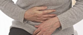
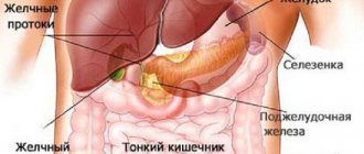
An elderly citizen very often faces a difficult situation when it is very painful or completely impossible to go to the toilet. Pathology of the digestive tract is a consequence of concomitant diseases, cancer. We will tell you the causes and symptoms of the disease and what to do in case of intestinal obstruction in older people.
Select a service for your relative | |||
| Caring for bedridden people | Disability care | Elderly care | Care after a heart attack |
Obturation, what is it?
Constipation is a blockage of the genital anatomical formation with a violation of its patency. The patient is no longer helped by strong laxatives, enemas, or dietary nutrition. When going to the toilet he experiences severe pain. In addition, with cancer, the stool has a liquid consistency with bloody discharge. Then you should pay close attention to your well-being and urgently contact a medical facility.
Types of pathology
Young people with this problem rarely go to doctors. People of advanced age suffer from deterioration of peristalsis or improper functioning of blood circulation. The condition of impaired movement of contents during bowel movements is a consequence of reduced mobility and long-term disorders. There are three forms of obstruction:
- Dynamic. The variety can be paralytic (myocyte activity decreases) and spastic (intestinal tone increases). It occurs in a chronic form or has an acute nature, which often leads to death.
- Vascular occurs due to poor blood circulation (for example, heart attack). Both types of intestines become blocked.
- Mechanical. Occurs after surgical intervention. The resulting adhesions can pinch the intestines, be compressed by internal tumors or a vertebral hernia. Often, constipation occurs due to the occurrence of prostate adenoma, which can completely block the passage. The entry of a foreign body also clogs the passage.
Constipation is the main symptom of intestinal obstruction in older people
Obstipation is manifested by difficult or insufficient defecation (feces). The frequency of bowel movements does not exceed two times in seven days. In severe cases, bowel movements occur 2-3 times a month.
Phases of pathology
There are three degrees of the course of OKN:
- "Ileus scream." From the moment the intestinal hemocirculation begins until complete emptying, about 12-14 hours pass. At this time, the eruption occurs with acute pain and muscle spasms.
- Intoxication of the body is accompanied by bloating and decreased gas production. Painful sensations weaken, the cramping nature stops, and they become aching. Emptying occurs after one and a half days. The body is experiencing a lack of water.
- Peritonitis occurs after a person has not gone to the toilet for 36 hours. Inflammation of the peritoneal layers occurs. If timely assistance is not provided, death may occur.
Consequences after self-medication
Therapy should only take place under the supervision of a doctor, that is, in a hospital. Home treatment will not bring positive results, since it is necessary to adhere to conservative therapy, which includes:
- intravenous drips that maintain water-salt balance;
- drugs to normalize peristalsis;
- antispasmodics for pain relief;
- laxatives;
- enemas, where a probe is inserted through the mouth.
If a person ignores conservative treatment, subsequent complications can lead him to the operating table. Part of the organ is removed and a bypass anastomosis is formed.
Intussusception in adults is a type of intestinal obstruction, consisting in the introduction of one intestinal segment into the lumen of another with compression of the mesentery, observed in less than 5% of all patients with intestinal obstruction [1-3]. Intussusception in adults is most often caused by organic intestinal lesions (81.8% benign, 18.2% malignant) [1, 4]. Malignant neoplasms account for 66% of colonic and 30% of small intestinal intussusceptions [1, 3]. Adenocarcinoma is the most common cause of colonic intussusception, and metastatic disease is the most common cause of small intestinal intussusception [5]. The most common ileocecal invaginations caused by a benign tumor are: leiomyoma, adenoma, lipoma, hamartoma of Brunner's glands, hemangioma, adenomyoma, neurofibroma, desmoid tumors [6, 7]. Other factors predisposing to intussusception are anorexia, malabsorption, impaired intestinal wall tone, unregulated anticoagulant therapy, causing submucosal hemorrhages and hematomas [1, 8].
The majority of intussusceptions in adult patients occur in the small intestine (75–80%) and are benign in nature [9]. The clinical picture of intussusception is most often nonspecific and corresponds to the phenomena of small or large intestinal obstruction. The most common symptoms are abdominal pain, nausea, vomiting, lack of stool, gastrointestinal bleeding, and bloating [8].
The problem of diagnosing and treating intussusception remains relevant due to the low incidence, nonspecific manifestations and the need for quick decision-making. In emergency patients presenting with clinical manifestations of acute abdominal pain, intussusception should be considered in the differential diagnosis. Surgery remains the preferred treatment option in adults. Taking into account the fact that an organic lesion can be malignant, even in emergency surgery, the principles of oncological radicalism should be observed.
We present 3 clinical observations of the development of intestinal obstruction with small-colic intussusception during the period from June 2016 to August 2021. These clinical observations indicate the difficulties of diagnosis, the possibilities of X-ray examination using contrast, the use of ultrasound in the dynamics of treatment and the favorable outcome of surgical intervention in elderly patients.
Patient E
., 30 years old, was admitted on the 3rd day from the onset of the disease with complaints of cramping pain in the lower and left parts of the abdomen, nausea, repeated vomiting, delayed passage of stool and gas, general weakness. From the collected medical history, it is known that the patient underwent a caesarean section 8 years ago.
Upon examination in the emergency department: a moderately swollen abdomen, soft on palpation, painful in the peri-umbilical region, along the midline of which, slightly to the left of the navel, a volumetric formation measuring 7x7 cm, soft-elastic consistency, mobile, moderately painful on palpation is determined. Peristalsis is auscultated and weakened.
X-ray examination of the abdominal cavity: on the left in the mesogastrium there are single pneumatized loops of the small intestine, without fluid levels, their lumen is not expanded; in the left parts of the colon there is a moderate amount of gas, against the background of which it is impossible to exclude an additional formation with a convex contour in the lumen of the transverse colon at level LII-III (Fig. 1).
Rice. 1. General radiograph of the abdominal organs (upon admission).
Ultrasound of the abdominal cavity: in the area of the right bend of the ascending colon there is a deformed fragment of the small intestine (like intussusception) in the form of a four-layer structure over 6 cm, with a diameter of up to 4 cm, the wall of which is thickened to 0.45 cm, with edematous folds. In digital Doppler mapping mode, blood flow is recorded in the wall. In the hypogastrium, the separation of the peritoneal layers between the loops is up to 0.4 cm, the contents are anechoic (Fig. 2).
Rice. 2. Ultrasound picture of the abdominal organs (the arrow indicates the intussusception).
The patient was hospitalized in the surgical department, where conservative treatment was performed (nasogastric intubation, infusion-spasmolytic therapy, cleansing enema) with some positive effect in the form of pain reduction, cessation of vomiting, and dynamic X-ray monitoring was performed.
During the assessment of the passage of a water-soluble contrast agent along the gastrointestinal tract on the left at level LIII-IV, against the background of a small amount of gas in the projection of the left flexure of the colon, an intraluminal filling defect of a round shape with a diameter of at least 5 cm was detected. During the control study after 2 hours 30 minutes, a clearly visible the contrast agent does not enter the lumen of the right parts of the colon. At the level of the above-described formation, there is an accumulation of contrast agent in the form of a series of concentrically located rings. The changes most likely correspond to intussusception of the small intestine into the colon, with the head of the intussusception located at the level of the left sections of the transverse colon.
Despite conservative treatment, the patient’s condition worsened: abdominal pain intensified and nausea persisted. Taking into account the negative dynamics of the clinical and instrumental picture of the disease, 6 hours after admission, emergency video laparoscopy was performed: during inspection of the abdominal cavity, a small amount of serous effusion in the pelvic cavity; the terminal section of the ileum (for about 80 cm) is dilated to 3 cm, there is gas and liquid contents in the lumen; in the subhepatic space, a small intestinal intussusception was visualized into the cecum, measuring 8x5 cm in the area of the ileocecal junction; Disinvagination of the small intestine was performed, intestinal obstruction was eliminated, and the abdominal cavity was drained.
On the 5th day after surgery, a colonoscopy was performed. In the ascending colon, an exophytic submucosal formation 10-11 cm long is determined, with a smooth, unchanged mucous membrane, emanating from the medial wall, almost completely blocking the intestinal lumen; on the surface of the formation an ulcerative defect measuring 0.8x1.5 cm with light dense fibrin is visualized at the bottom. Histological examination showed: the formation is represented by the mucous membrane of the colon with an ulcerated area extending to the muscular lamina propria, the ulceration area is lined with granulation tissue permeated with inflammatory infiltrate, the edges are epithelialized, and purulent-necrotic detritus is adjacent to the surface. PAS staining revealed no tumor mucus-forming cells.
Taking into account the presence of a formation in the colon and the threat of repeated intussusception, on the 6th day after the first operation, relaparoscopy, video-laparoscopic right hemicolonectomy, and drainage of the abdominal cavity were performed.
Histological examination with Van Gieson staining showed: the tumor is represented by chaotically intertwined bundles of smooth muscle cells of different sizes, with a pronounced vascular component, an extremely scanty admixture of collagen fibers, and focal circulatory disorders. Lymph nodes with intact structure, without tumor lesions. Conclusion: submucosal leiomyoma of the colon (Fig. 3).
Rice. 3. Macropreparation. Arrows indicate colon leiomyoma (intraoperative photograph).
The postoperative period proceeded smoothly. In satisfactory condition, the patient was discharged under observation by a surgeon and oncologist at her place of residence.
Patient P.
, 90 years old, was admitted with complaints of cramping abdominal pain, nausea, vomiting, and delayed stool passage for 3 days.
From the anamnesis it is known that at the age of 60 the patient underwent supravaginal amputation of the uterus without appendages, and at the age of 76 she underwent videolaparoscopic cholecystectomy.
Upon examination in the emergency department, the patient's condition was moderate. The tongue is dry, covered with a white coating. The abdomen is swollen, soft on palpation, painful in the meso- and hypogastrium. No bowel sounds are heard. The symptom “splashing noise” in the mesogastrium is determined.
During a survey X-ray examination of the abdominal organs: the stomach is moderately pneumatized, with a wide level of fluid in the lumen; on both sides, multiple pneumatized loops of the small intestine are identified with horizontal levels of fluid in the lumen with the formation of small intestinal arches, small levels of fluid are noted on the left in the lower sections - the “string of beads” symptom; the lumen of the small intestine is dilated to 4.5 cm, the large intestine does not contain gas. Conclusion: small intestinal obstruction.
Ultrasound of the abdominal cavity: echo signs of free fluid interloop (up to 3 cm in the right, up to 1.8 cm in the left).
The patient was hospitalized in the surgical department, where conservative treatment was performed (cleansing enema, nasogastric intubation, intravenous infusion of crystalloid solutions and antispasmodics). As a result, the abdominal pain decreased, gases passed, and stools of normal color and consistency.
An X-ray examination was performed to assess the passage of a water-soluble contrast agent through the gastrointestinal tract. After 2 hours from the start of the study, part of the contrast agent is detected in the stomach, part - in the small intestine. After 4 hours from the start of the study, further movement of the contrast agent through the small intestine to the distal sections is noted. After 6 hours, the contrast agent is detected in the small intestine along its entire length, after 8 hours - in the large intestine along its entire length. Conclusion: passage through the intestines is preserved, but the timing is slowed down.
To exclude mechanical intestinal obstruction and assess the condition of the right parts of the colon, a contrast enema was performed. On a plain X-ray of the abdominal cavity before the study, multiple pneumatized loops of the small intestine are identified, dilated to 4.5 cm, with multiple horizontal levels of fluid in the lumen, with the formation of multiple small intestinal arches, in the projection of the small pelvis, loops of the small intestine filled with fluid; a moderate amount of gas and liquid contents along the entire length of the colon. In the process of a contrast study, in the projection of the ascending colon, a large oblong filling defect is determined, at least 10 cm in length, with a polycyclic contour, which is not visible on the intestinal contour during a polypositional study - intussusception (?), exophytic formation (?). Throughout the rest of the colon, the contours of the colon are clear, smooth, the walls are elastic, and haustration is preserved. Bowel emptying is incomplete.
To clarify the nature of changes in the area of the ileocecal junction identified during a contrast enema, the next day per os
A cooled suspension of barium sulfate in an amount of 100 ml is given. During a control study 8 hours after taking a contrast agent, the terminal ileum is contrasted; the right parts of the colon - the cecum and ascending colon are not contrasted. In the projection of the right sections of the transverse colon, an oval filling defect is determined. The contrast agent enters the lumen of the colon in small portions immediately into the right sections of the transverse colon. The lumen of the ileum is deformed, the diameter is unevenly narrowed from 1.5 to 0.6 cm over a length of at least 9 cm. In the projection of the right sections of the transverse colon, there is an oval-shaped filling defect - the head of the intussusception, which was determined during a contrast enema in the projection of the ascending colon. Conclusion based on the results of two contrast studies: radiological signs of intussusception of the terminal ileum into the colon - to the right sections of the transverse colon.
Due to the persistent clinical and instrumental picture of small intestinal obstruction, a laparotomy was performed 72 hours after admission. During inspection: the small intestine is about 3 cm in diameter, contains gas and liquid intestinal contents; palpation in the lumen of the cecum reveals an invaginated terminal portion of the ileum, in which a dense tumor-like formation with a diameter of up to 4 cm is palpated; 20 cm from the intussusception in the lumen of the small intestine there is a dense tumor-like formation with a diameter of about 2 cm (Fig. 4).
Rice. 4. The site of intussusception is indicated by an arrow (intraoperative photograph). A right hemicolonectomy was performed with removal of 30 cm of the ileum, including the tumor (Fig. 5).
Rice. 5. Macropreparation. Malignant melanoma, indicated by an arrow (intraoperative photograph).
Pathohistological conclusion: foci of proliferation of a neuroendocrine tumor (carcinoid?) in the wall of the small intestine.
Immunohistochemical conclusion: the immunophenotype features of the tumor correspond to malignant melanoma.
The postoperative period proceeded without complications. The patient was discharged in satisfactory condition under observation by a surgeon and oncologist at her place of residence.
Patient Z.
, 79 years old, was admitted 9 hours after the onset of the disease with complaints of abdominal pain, more in the right side, nausea, and single vomiting.
From the anamnesis: extirpation of the uterus and appendages was performed.
When examined in the emergency department, the condition was of moderate severity. The tongue is dry. The abdomen is moderately swollen, soft on palpation, painful in the upper and right parts of the abdomen along the colon. Peritoneal symptoms are negative. Peristalsis is preserved. There are no dysuric disorders. Urine is straw-yellow.
Ultrasound of the abdominal cavity: echo signs of a space-occupying formation in the pelvic cavity; in the projection of the terminal ileum, the dome of the cecum, a fragment of intestine 7.8×4 cm with a wall thickness of 1 cm and reduced echogenicity is identified more to the right.
During a general X-ray examination of the abdominal cavity: small intestinal arches and fluid levels are noted on both sides; the small intestine is dilated to 3.5 cm, with edematous folds of the mucous membrane and intestinal walls, there is almost no gas in the large intestine.
Taking into account the clinical and instrumental picture of intestinal obstruction, the patient was offered emergency surgical intervention - diagnostic video laparoscopy, which the patient refused.
30 hours after admission, a repeated X-ray examination: to the left and right of the spine, more to the left, small intestinal arches and unclear fluid levels are noted - the “string of beads” symptom, the small intestine is dilated to 3.5 cm, with edematous folds and walls; There is almost no gas in the colon.
Taking into account the negative dynamics, the patient was again offered emergency surgery. Consent has been received. A midline laparotomy was performed: during inspection of the abdominal cavity there was a small amount of serous effusion, the loops of the small intestine were dilated to 4 cm, hyperemic, peristalsis was observed, vascular pulsation was clear, nasointestinal intubation was performed, in the area of the ileocecal junction there was intussusception of the ileum into the cecum (Fig. 6) ;
Rice. 6. Intussusception of the ileum into the cecum (indicated by an arrow), areas of necrosis (intraoperative photograph). after straightening the intussusception, focal necrosis of the invaginated ileum and its perforation were discovered, and therefore a right hemicolectomy was performed with the removal of 30 cm of the ileum, a side-to-side ileotransverse anastomosis was formed, and the abdominal cavity was sanitation and drainage.
Pathohistological conclusion: intussusception of the small intestine with necrosis throughout the entire thickness of the wall. No pathological formations were found in the removed specimen.
In the postoperative period, infusion-transfusion, antibacterial, antispasmodic, and symptomatic therapy was carried out with a positive effect. The postoperative period was without complications. She was discharged in satisfactory condition under the supervision of a surgeon at her place of residence.
20 days after discharge, a contrast X-ray examination of the colon was performed - a contrast enema. X-ray report: operated colon; condition after right hemicolonectomy with normal function of ileotransverse anastomosis.
Thus, the clinical picture of small-colic intussusception is manifested by intestinal obstruction, most often low-grade small-bowel obstruction. X-ray examination using contrast, carried out in parallel with the preoperative preparation of the patient (infusion therapy, nasointestinal intubation, siphon enemas), can improve the quality and timeliness of diagnosis of intussusception. With the introduction of endosurgical technologies into clinical practice, the possibilities for using low-traumatic laparoscopic interventions both to diagnose the cause and to eliminate intestinal obstruction are increasing.
The authors declare no conflict of interest.
*e-mail; https://orcid.org/0000-0002-5717-6520
How acute and chronic obstruction develops
The acute phase is characterized by nausea and vomiting. At the initial stage, vomiting occurs with the contents of the stomach, so far without stagnation. The disease is aggravated by loss of fluid (water) and electrolytes. It occurs without a typical pain syndrome; cramping pain occurs only in 30% of the population suffering from acute insufficiency. Retention of stool with continued passage of gas does not occur in everyone.
After a long period, a phase of “imaginary well-being” begins, during which contractions and vomiting stop. Acute pain is replaced by aching, non-localized and dull. At first, the patient feels relief, but soon the stomach becomes very swollen. Intoxication occurs when “fecaloid” vomiting returns with putrid contents resembling feces. Peritonitis then develops. This is due to a slower passage through the intestinal tract. That is, bowel movements occur 2-3 times a week. In addition, there is a slight asymmetrical swelling of the abdomen and cramping pain.
Leave a request for selection of a boarding house
for an elderly person with intestinal obstruction
Causes of intestinal obstruction in older people
The main etiological factors include:
- The appearance of adhesions after surgery.
- Concomitant diseases: acute cholecystitis, inflammation of the appendage of the cecum, pancreatitis, etc.
- Taking certain medications, such as anesthesia medications, opiates.
- The appearance of twists and knots.
- Malignant and benign neoplasms.
- Hernia development.
- Formation of sand and stones in the gall bladder.
- Entry of foreign objects into the gastrointestinal tract.
- Infestation with parasites.
- Sedentary lifestyle.
- Constant diets.
- Sedentary work.
- Dietary fiber can cause a lump to form.
Bruises, bruises, kidney failure, pleurisy, cracked or broken ribs, inflammation of the lungs and pancreas, damage to the central nervous system (strokes, spinal column injuries and skull damage) can also lead to serious illness.
How does pathology manifest itself?
Clinical symptoms look like this:
- Delay in defecation for 36 hours or more.
- The appearance of vomit. Initially, they are caused reflexively and contain food mass that is indigestible by the stomach. At the last stage of the disease, vomiting takes on a yellow-green tint.
- Pain in the peritoneum can be cramping, sharp and aching. If assistance is not provided in a timely manner, a person may lose consciousness from pain.
- The occurrence of flatulence.
- Constipation. This phenomenon can confuse a person, since on the first day he has stool. But the pain and nausea reflex do not stop. After a rectal examination, it is revealed that stagnation of feces has occurred in the small intestine. This leads to the absorption of rotting products and, as a result, to intoxication of the body.
Symptoms
Intussusception in children of the first year of life often manifests itself as cramping abdominal pain that occurs every 10-20 minutes. In later life, the pain is constant. Due to hemorrhages in the mucous membrane, blood is found in the stool. Sometimes the stool takes on the appearance of “raspberry jelly.” Palpation reveals a mobile, painful formation that changes its shape and location depending on the duration of the disease. Body temperature, as a rule, remains within normal limits. In severe cases, pronounced signs of intoxication appear.
In children of the first year of life, intussusception is manifested by cramping pain in the abdomen
In addition, intussusception may be accompanied by the following symptoms:
- vomit;
- retraction of the abdomen;
- defecation disorders;
- prolapse of intussusception through the anus;
- relaxation of the anal sphincter.
Establishing diagnosis
When a citizen applies to a medical institution, the doctor immediately prescribes an examination:
- Performs abdominal palpation.
- Refers for x-ray examination.
- Blood is taken and sent for analysis.
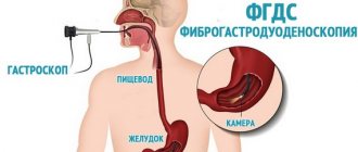
- The colon and rectum are examined with an endoscope.
- Irrigoscopy is performed using the method of retrograde injection of the drug to obtain an image.
- Examine with ultrasound.
- Carry out computer diagnostics.
Often a rectal examination is used first. Women are scheduled to visit a gynecologist. If the clinic is equipped with the necessary equipment, then they will send you for a survey radiography. With the introduction of a contrast agent, the localization of pathological processes can be accurately determined. Since the disease is often caused by parasites, the feces are examined for the presence of eggs of various helminths.
How to treat intestinal obstruction in older people
Taking any independent action is strictly prohibited! Giving an enema and taking painkillers will not lead to a positive result; they can worsen the situation. You need to urgently go to the hospital for help. They will empty the abdominal cavity with a probe by inserting it into the mouth or nose. If necessary, antispasmodics are prescribed, a novocaine blockade is performed, and a siphon enema is administered.
If droppers with saline solutions do not help (this indicates a mechanical type of pathology), then an emergency operation is performed.
What should relatives do at the first symptoms of the disease in elderly people?
Place the patient on a comfortable bed and place a basin near him to collect the vomit. Call an ambulance, do not take any further action. Do not give painkillers or laxatives under any circumstances.
Causes of constipation
The movement of food occurs due to contraction of the intestinal walls. Impaired peristalsis may be accompanied by relaxation of the muscle layer or prolonged spasms. The cause of the onset of the disease may be:
- surgical intervention;
- taking medications;
- accompanying illnesses;
- formation of nodules, twists;
- tumors;
- hernias;
- the presence of stones in the gall bladder;
- entry of foreign objects;
- bruises, hematomas;
- rib fractures;
- problems with blood vessels;
- traumatic brain injuries;
- presence of helminths.
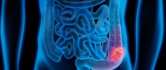
Symptoms of intestinal obstruction in older people
An elderly person begins to complain of pain in the abdomen, first points to a certain area, then begins to feel painful spasms throughout the entire volume of the peritoneum. At this time, nausea begins, and later vomiting appears. Trying to go to the toilet leads nowhere. On the contrary, diarrhea may develop. The worst thing is if it occurs with blood inclusions. The face and body are covered with cold sweat, the skin becomes noticeably pale, and the person assumes a “fetal” position. He may lose consciousness from pain and intoxication. In such cases, he urgently needs to be taken to the hospital or an ambulance is called. Most likely he will have surgery.
Causes of intestinal obstruction
Most often, surgeons have to deal with intestinal obstruction caused by malignant tumors and polyps, hernias, and adhesions in the abdominal cavity. Other reasons less common:
- Diverticulitis is an inflammation of a diverticulum, a protrusion on the wall of the intestine in the form of a pouch.
- Gallstones that “fall through” into the intestines.
- Foreign bodies.
- Crohn's disease.
- Parasites are roundworms.
- Intestinal volvulus.
- Intussusception is a condition in which one part of the intestine invades another. In adults, this pathology occurs rarely; it can be caused by malignant tumors.
Our doctors will help you
Leave your phone number
Diagnostics
Upon arrival at the medical facility, the patient is immediately prescribed an examination, where the following manipulations are performed:
- take blood for analysis;
- feces are passed;
- palpation and rectal examination are performed;
- X-rays are taken;
- conduct research using ultrasound, computer diagnostics, and endoscope;
- perform irrigoscopy measures.
Conservative treatment method
After diagnosis, gastrointestinal tract lavage is prescribed by inserting a probe through the mouth or nose. To relieve pain, injections containing analgesics and antispasmodics are given. To normalize peristalsis in the paralytic form of the disease, Proserin is used. A novocaine blockade is often performed. To maintain the water-salt balance of the body, droppers are placed.
Preventive actions
For an elderly person, it is very important to engage in feasible physical labor, spend less time in bed and move more. This could be: light gymnastics, walking, weeding garden beds, etc.
In addition, he must eat properly and follow a diet. It is necessary to exclude smoked meats, pickles and marinades, spicy and heavy foods from the diet. You should drink at least 1.5 liters of water per day. You should be careful about lifting weights; at this age, a bag weighing 2-3 kg is allowed. Regular examination by doctors will help identify various diseases and the presence of parasites that contribute to the development of constipation.
How to get rid of fecal stones?
Enema
Enema is one of the effective ways to combat fecal stones. The procedure can be carried out in a hospital or at home. This is acceptable if the patient’s condition does not require urgent medical intervention, that is, there are no signs of complete obstruction of the intestinal lumen.
It is recommended to start treatment with a cleansing enema. To do this you need:
- Prepare a solution for cleansing - it is advisable to use a herbal decoction, which is prepared at the rate of 1 tablespoon of herbal mixture (chamomile, dill, celandine) per 0.5 liters of boiling water.
- Leave the resulting solution for about half an hour.
- Pour it into a syringe.
- Place the patient on his side, with his legs pulled toward his stomach.
- Insert the tip of the syringe into the patient's anus.
- Slowly introduce the solution into the intestines.
- Wait 10-15 minutes, after which the patient should go to the toilet.
If a conventional cleansing enema is ineffective, a more extensive intervention is performed - a siphon enema. It involves repeated administration and removal of fluid. 1-2 liters are administered at a time, the total volume of solution used for the procedure is 15-20 liters.
Hydrocolonoscopy
Hydrocolonoscopy is a therapeutic intervention, which is a type of enema therapy. The procedure is carried out using special dialysis equipment, which allows for flow-through rinsing of the intestines with water or special solutions. The total volume of liquid used is approximately 20-30 liters. The duration of each session is only 20-40 minutes. The intervention can be performed on an outpatient basis; hospitalization of the patient is not required.
Repeated colon cleansing allows you to remove small fecal stones from the digestive system. Colon hydrotherapy is carried out in several sessions, the interval between which is 2-3 days. Repeated procedures ensure that all coprolites are removed from the intestines.
Laxatives
Laxatives are a fairly effective method of combating coprolites, but they have a number of limitations. Drugs from this group may have different mechanisms of action. This can affect the patient's condition and even worsen the course of his disease.
In addition, in most cases, the use of laxatives is accompanied by the removal of important electrolytes from the body, which also has certain consequences. For example, a decrease in potassium content worsens the course of cardiovascular pathologies.
Therefore, to prevent adverse consequences, self-medication with laxatives is strictly prohibited! The medications should be selected by the attending physician after a complete examination of the patient.
| A drug | Group of laxatives | Reception features |
| Castor oil | Agents that stimulate peristalsis | 1 tablespoon twice a day, it is possible to use oil in capsules |
| Sodium sulfate | Drugs that help stretch the intestinal walls | In enemas 120 ml per procedure |
| Lactulose | Stool thinners | 2-3 tablespoons of the product in the first 3 days, after – 1-2 tablespoons |
Diet
The diet for coprolites should comply with several general principles that help restore normal peristalsis. These include:
- Fractional meals. You need to eat 5-6 times a day, in small portions.
- Gentle nutrition. Food should cause minimal discomfort to the intestines. This applies not only to the chemical composition of products, but also to temperature characteristics. Too hot foods or drinks, as well as too cold foods, are prohibited.
- Balanced diet. The patient's diet should include sufficient amounts of nutrients, vitamins, and electrolytes.
How doctors examine a patient
When diagnosing, professionals adhere to the following principles:
- Blood is taken during intoxication.
- You can bring feces and other tests with you from home during an exacerbation or take them in the hospital.
- Before radiography, it is necessary to empty the gastrointestinal tract.
All manipulations are carried out only with the permission of the patient or his guardian.
Treatment
The elderly person is left in a hospital bed until he fully recovers. At this time, pain is relieved with the help of analgesics and antispasmodics. If necessary, a blockade is carried out with Novocaine and siphon enemas are given. If such actions do not lead to a positive result, then he is prepared for surgery.
Surgery for intestinal obstruction in the elderly
Removing obstacles occurs as follows:
- The abdominal cavity is opened.
- The contents of the afferent sections of the intestine are removed.
- Resection is performed.
- Bypass anastomoses are formed.
- I apply fistulas.
The outcome of the operation may vary; successful recovery depends on the rehabilitation period.
Treatment of obstruction
The main method of treatment for patients is surgical intervention. If intestinal obstruction is suspected, the patient is urgently hospitalized. Conservative therapy is ineffective in the presence of paresis. Until specialists have established an accurate diagnosis, it is unacceptable to give the patient antispasmodics and give an enema.
In a hospital setting, the patient's stomach is emptied using a tube, and the intestines are emptied in the same way. The probe is inserted through the nasal cavity; if necessary, doctors can perform a siphon enema. After this, the attending physician gives the patient an antispasmodic for pain relief.
If paralytic intestinal obstruction is detected, specialists use Proserin, which normalizes intestinal motor functions. If necessary, the patient will undergo a novocaine blockade. In the future, doctors treat the patient according to the following scheme:
What does an MRI of the intestine show?
- Intravenous administration of saline solutions is carried out. If nothing has helped so far, we can talk about the presence of a mechanical type of intestinal obstruction. In this situation, urgent surgical intervention is performed.
- Part of the intestine is removed when necrosis is established. This type of surgery is called resection.
- The specialist very carefully applies anastomoses and proceeds to eliminate the intussusception.
- If a volvulus is noted, the surgeon urgently untwists the intestine and fixes it. A temporary colostomy may be required. With the development of peritonitis, it is necessary to perform a transversostomy, during which an external fistula will begin to form in part of the transverse colon. If a patient's blood pressure drops sharply, he is prescribed Dopamine or Dobutamine.
Alas, not everyone understands how dangerous intestinal obstruction is and how to help a sick elderly person. Having normalized peristalsis or eliminated obstruction, it is necessary to urgently switch to infusion therapy and start taking antibiotics. The acute form of the disease can lead to the death of the patient, so when the first symptoms of a dangerous disease appear, it is important to immediately seek help from a medical specialist.
Intestinal obstruction requires urgent hospitalization
Keeping a diet
After the surgical procedure, the patient is not allowed to drink or eat for 12 hours. Then he is supported by droppers with nutrient solutions injected directly into the intestine. After a few days, the mixture is given to eat through a tube.
When the patient is able to fully consume normal food, he is allowed fermented milk products, soups cooked in water without adding fat, and baby food. He must follow diet No. 4, where all dishes are cooked boiled or steamed with a minimum amount of salt.
Menu
The diet is determined by the attending physician and is observed for three to four weeks after surgery. A person can eat:
- White meat chicken and turkey pate.
- Kefir, fermented baked milk, yogurt, cottage cheese.
- Vegetables and fruits chopped in a blender.
- Liquid in unlimited quantities. Only mineral or regular water should be alternated with beet, carrot, apple, peach and apricot juices.
Fried, smoked, grilled and fatty foods are strictly prohibited. Pepper and other spices, strong-smelling herbs, acidic foods (lemon, cranberry, orange, tangerine, etc.) are excluded from the diet. After the operation, you should not drink alcoholic beverages, tea, coffee or carbonated mineral water!
Causes of pathological intestinal disorders
The normal functioning of the absorptive organ of the gastrointestinal tract is influenced by a large number of factors, both external and internal. But where does the original source of the development of illnesses come from? Nikolai Amosov argued: “All human illnesses and misfortunes arise from poor nutrition. We are what we eat." There is a great deal of truth in this expression. After all, preservatives, nitrates, pesticides and various heavy metals settle in the sections of the colon. Excessive accumulation of these structural components affects the normal metabolism and functioning of cells and tissues. A person causes himself a lot of problems by eating on the go and chewing food poorly. This can also include abuse of alcohol and various medications. The causes of slagged and clogged intestines are as follows:
- High calorie foods.
- Food that has been prepared through prolonged heat treatment (canned food, juices, casseroles).
- Insufficient amount of fluid in the human body.
- Weak motor activity (lying down lifestyle).
- Psychological disorders and stressful moments.
The above reasons can be safely supplemented by unsystematic bowel movements. Healthy people always monitor the frequency of this “ritual” and control the physiological activity of the body. Medicine says that the most favorable time to visit the toilet is early morning and evening. In the absence of a clear pattern of defecation, feces stagnate in the bends and folds of the suction organ. The malignant consistency gradually thickens and transforms into fecal stones. Thus, the nerve endings of the intestine stop promptly responding to the pressure of decay products. In practice, a person does not feel the urge to go to the toilet. What follows is a closed cycle. Poor quality nutrition develops into clogging of all parts of the intestines, which leads to atrophy of special impulses. As a result, chronic atony and intoxication are formed, which complicates the stagnation of the absorption organ. It becomes impossible to go to the toilet.
Experts say that the total mass of stones (feces) in the intestines can reach from 3 to 5 kg. Both thin and fat people can equally face this situation.