brief information
The content of the article:
1.1 Definition
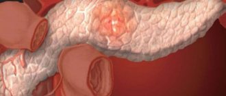
Acute pancreatitis (AP) is an initially aseptic inflammation of the pancreas, which may damage surrounding tissues and distant organs.
1.2 Etiology and pathogenesis
Etiological forms of pancreatitis:
- Acute alcohol-nutritional – 55%
- Acute biliary – 35%
- Acute traumatic 2 – 4%
- Other etiological forms – 6 – 8%
Factors of aggression
Primary:
- trypsin, chymotrypsin cause proteolysis of proteins;
- phospholipase A2 destroys cell membranes;
- lipase leads to lipolytic necrosis in the gland, fiber and mesentery of the intestine;
- elastase destroys the vascular wall and connective tissue structures, which leads to necrosis.
Secondary:
- activation of the kallikrein–kinin system by enzymes disrupts microcirculation.
Tertiary:
- macrophages, mononuclear cells, neutrophils produce cytokines that suppress immunity.
Quaternary:
- cytokines, enzymes, metabolites increase the permeability of the intestinal wall, which facilitates the entry of toxins into the bloodstream and lymph and damage target organs.
Factors of aggression and organ dysfunction create a syndrome of “mutual burden.”
Phases of acute pancreatitis:
- Edematous (interstitial) pancreatitis 80-85% is mild with the rare development of local complications or systemic disorders, has no phases.
- Necrotizing pancreatitis (pancreatic necrosis) in 15-20%, moderate or severe, phase course with 2 peaks of mortality - early (1 week each of phases IA and IB) and late (weeks and months).
Phase I A – up to 3 days, the formation of foci of necrosis in the parenchyma and the development of endotoxemia with multiple organ failure.
Phase I B – the body’s reaction to foci of necrosis with resorptive fever and the formation of a peripancreatic infiltrate.
Phase II – aseptic and septic sequestration.
1.3 Epidemiology
- Prevalence 32-389 per 1 million population.
- Mortality rate is 6-12 per 1 million population.
- Overall mortality 2.5%-3.5%; postoperative 20%-25%
- Since 2009, in the structure of “acute abdomen” AP has moved from 1st place to 2nd place with a share of 25%-35%, giving way to acute appendicitis.
1.4. Coding according to ICD-10
Acute pancreatitis (K85):
- K85.0 – Idiopathic acute pancreatitis;
- K85.1 – Biliary acute pancreatitis: cholelithiasis pancreatitis;
- K85.2 – Alcoholic acute pancreatitis;
- K85.3 – Drug-induced acute pancreatitis;
- K85.8 – Other types of acute pancreatitis;
- K85.9 – Acute pancreatitis, unspecified.
1.5 Classification
Mild AP – pancreatic necrosis does not form (edematous pancreatitis) and organ failure does not develop. Moderate AP - with one of the local manifestations (infiltrate, pseudocyst, abscess), or/and the development of transient - no more than 48 hours of organ failure. Severe AP - with either not limited infected pancreatic necrosis (purulent-necrotic parapancreatitis) and/or the development of persistent organ failure.
Diagnostics
Clinical manifestations depend on the morphological form, phase of the disease, the severity of the systemic inflammatory response syndrome and organ failure.
Each phase of the disease corresponds to a clinical and morphological form, so diagnosis is carried out depending on the phase of the disease.
Scale of criteria for primary express assessment of the severity of AP (St. Petersburg Research Institute of SP, 2006):
- peritoneal syndrome;
- oliguria (less than 250 ml in 12 hours);
- skin symptoms (facial hyperemia, marbling, cyanosis);
- SBP less than 100 mmHg;
- encephalopathy;
- Hb more than 160 g/l;
- leukocytes more than 14 x109/l;
- blood glucose more than 10 mmol/l;
- urea more than 12 mmol/l;
- metabolic disorders according to ECG;
- cherry or brown-black enzymatic exudate during laparoscopy/laparocentesis;
- during laparoscopy, widespread enzymatic parapancreatitis extending beyond the boundaries of the omental bursa and spreading along the flanks;
- common steatonecrosis during laparoscopy;
- lack of effect from basic therapy.
- Scale rating:
5 signs – with 95% probability a severe form of AP. 2-4 signs – moderate AP, hospitalization in the ICU. 0 – 1 sign – mild form of AP, hospitalization in the surgical department.
The SOFA scale is used to assess organ and multiple organ dysfunctions.
If it is impossible to determine the severity of AP using scales, clinical and laboratory criteria for AP are:
- signs of systemic inflammatory response syndrome (SIRS);
- hypocalcemia <1.2 mmol/l,
- hemoconcentration: Hb> 160 g/l or Ht> 40 units, glucose> 10 mmol/l;
- CRP >120 mg/l;
- shock
- renal failure (oligo-anuria, creatinine >177 µmol/l);
- liver failure (hyperfermentemia);
- cerebral insufficiency (delirium, stupor, coma);
- gastrointestinal bleeding (more than 500 ml/day);
- coagulopathy
Urgent EPST with lithoextraction for impaction of a stone in the major duodenal papilla (MDP):
- intense pain syndrome that is not relieved by narcotic analgesics;
- rapidly progressing jaundice;
- absence of bile in the duodenum during FGDS;
- signs of biliary hypertension on ultrasound.
Indications for CT/MSCT (MRI):
- uncertainty of diagnosis and differential diagnosis;
- the need to confirm severity using clinical prognostic features;
- lack of effect from conservative treatment.
Time frame for MSCT (MRI):
- for the diagnosis of pancreatic necrosis on days 4–14 of the disease;
- as the disease progresses;
- in the absence of treatment effect;
- to clarify the localization of foci of suppuration before drainage interventions.
The Balthazar CT pancreatitis severity index is not a mandatory study, but is used to predict the severity of the disease.
Algorithms for the management of patients with acute and chronic pancreatitis
Acute pancreatitis
Acute pancreatitis is an acute inflammation of the pancreas, manifested by pain in the upper abdomen and increased levels of pancreatic enzymes in the blood and urine, in which clinical and histological changes completely resolve after the action of the etiological factor ceases. Scheme 1. Signs of severe acute pancreatitis
Organ failure and/or Local complications
Poor early prognostic criteria
Signs of organ failure: Shock: systolic blood pressure < 90 mm Hg. Pulmonary insufficiency: PaO2<60 mmHg. Renal failure: creatinine > 2 mg% Gastrointestinal bleeding > 500 ml/day |
The main goals of therapy for acute pancreatitis are to prevent systemic complications of the disease, necrosis of the pancreas, and prevent infection during the development of necrosis. Main systemic complications
acute pancreatitis are respiratory and renal failure, hypotension. Treatment of systemic complications is largely based on the elimination of inflammatory mediators, in particular activated pancreatic enzymes:
- Suppression of pancreatic secretion of enzymes (H2-blockers, proton pump inhibitors, anticholinergic blockers, glucagon, calcitonin, 5-fluorouracil, somatostatin and its analogue octreotide).
- Removing inflammatory mediators from the circulation. Protease inhibitors (for example, aprotinin and gabexate) have no effect. The effectiveness of new drugs that can influence cytokines, lysosomal hydrolases, and reactive oxygen compounds is expected.
- Peritoneal lavage.
Removal of common bile duct stones also reduces the risk of systemic complications. ERCP is indicated in the first 2-3 days after hospitalization of a patient with biliary pancreatitis and signs of biliary sepsis or organ failure. Table 1. Ranson severity criteria for acute pancreatitis
| Index | Pancreatitis | |
| Alcoholic | Biliary | |
| On admission: | ||
| * age of the patient, years | > 55 | > 70 |
| * leukocytosis, mm3 | > 16,000 | > 18,000 |
| * serum glucose, mg% | > 200 | > 220 |
| *Serum LDH, IU | > 700 | > 400 |
| * Serum AST, IU | > 250 | > 250 |
| During the first 48 hours: | ||
| * decrease in hematocrit, % | > 10 | >10 |
| * increase in serum nitrogen, mg% | > 5 | > 2 |
| * calcium level, mg% | < 8 | < 8 |
| * Arterial blood PaO2, mm Hg. Art. | < 60 | — |
| * base deficiency, meq/l | > 4 | > 5 |
| * estimated loss (sequestration) of fluid, l | > 6 | > 4 |
Prevent the occurrence of pancreatic necrosis
allows active infusion therapy.
Infection of necrotic areas
is associated with the movement of bacteria from the colon.
In 75% of cases, infection occurs with Escherichia coli, Klebsiella
and other gram-negative bacteria, in 20% -
Staphylococcus
and
Streptococcus sp.
The effectiveness of antibiotics for preventing infection has not yet been definitively established. However, it is recommended to use antibiotics in patients with pancreatic necrosis who have developed organ failure and who are at high risk of developing infection. Imipinem, ofloxacin and ciprofloxacin penetrate the pancreatic tissue to the greatest extent. Treatment of patients with acute pancreatitis should be differentiated depending on the severity of the disease (Scheme 1). The Ranson criteria (Table 1) and APACHE-II (Scheme 2) are of greatest importance for assessing the severity of acute pancreatitis.
Table 2. Glasgow Coma Scale (GCS) Counts one score per category
| Verbal reaction | oriented | 5 |
| inhibited | 4 | |
| the answer is out of place | 3 | |
| slurred sounds | 2 | |
| No answer | 1 | |
| Motor reaction | executes commands | 5 |
| indicates the location of pain | 4 | |
| flexion response to pain | 3 | |
| subcortical movements | 2 | |
| extensor response to pain | 1 | |
| Eye reaction | spontaneous | 4 |
| per voice | 3 | |
| for pain | 2 | |
| No | 1 | |
| Total GCS: | ||
Treatment of mild acute pancreatitis
The course of acute pancreatitis is considered mild if favorable prognostic signs are determined and there are no systemic complications. Treatment consists of maintenance therapy. It is necessary to adequately compensate for the loss of circulating fluid as a result of vomiting and diaphoresis. The occurrence of hypovolemia can lead to transformation of the disease into necrotizing pancreatitis. To relieve pain, you can use narcotic analgesics (meperidine 50-100 mg intramuscularly every 3-4 hours or hydromorphone), including morphine. If an infection of the respiratory system, biliary or urinary tract develops, appropriate antibacterial therapy is carried out. Eating is resumed on the 3-7th day of hospital stay after the disappearance of abdominal resistance, significant subsidence of pain, restoration of peristaltic noises and the appearance of a feeling of hunger in the patient. It is advisable to start with small meals consisting primarily of carbohydrates, which stimulate pancreatic secretion to a lesser extent than fats and proteins.
Drug treatment of severe pancreatitis
Patients with severe pancreatitis have a much higher risk of developing necrotizing pancreatitis, which can be determined by performing a CT scan with the introduction of a contrast agent. If the prognosis is unfavorable and signs of organ failure appear, the patient must be transferred to an intensive care unit for joint observation by a team of gastroenterologists, pulmonologists, surgeons and radiologists. Scheme 2. APACHE-II severity criteria for acute pancreatitis
A. Indicator of acute oridiological disorders
| above normal | below normal | ||||||||
| Physiological indicators | +4 | +3 | +2 | +1 | 0 | +1 | +2 | +3 | +4 |
| 1. Rectal temperature, °C | >41 | 39 — 40,9 | 38,5 — 38,9 | 36 — 38,4 | 34 — 35,9 | 32 — 33,9 | 30 — 31,9 | < 29,9 | |
| 2. Average blood pressure, mm Hg. | >160 | 130-159 | 110-129 | 70-109 | 50-69 | <49 | |||
| 3. Heart rate | >180 | 140-179 | 110-139 | 70-109 | 55-69 | 40-54 | <39 | ||
| 4. Respiratory rate | >50 | 35-49 | 25-34 | 12-24 | 10-11 | 6-9 | <5 | ||
| (regardless of ventilation) | |||||||||
| 5. Oxygenation A-aDO2 or PaO2 (mmHg) a FIO2 < 0.5 A-aDO2 value | >500 | 350-499 | 200-349 | <200 | |||||
| b FIO2 < 0.5 PaO2 only | PO2 >70 | PO2 61-70 | PO2 55-60 | PO2 <55 | |||||
| 6. Arterial blood pH | >7,7 | 7,6-7,69 | 7,5-7,59 | 7,33-7,49 | 7,25-7,32 | 7,15-7,24 | <7,15 | ||
| 7. Serum Na+, mmol/l | >180 | 160-179 | 155-159 | 150-154 | 130-149 | 120-129 | 111-119 | <110 | |
| 8. Serum K+, mmol/l | >7 | 6-69 | 55-59 | 35-54 | 3-34 | 25-29 | <25 | ||
| 9. Serum creatinine, mg% | >3,5 | 2-3,4 | 1,5-1,9 | 0,6-1,4 | <0,6 | ||||
| (Double value for acute renal failure) | |||||||||
| 10. Hematocrit, % | >60 | 50-59,9 | 46-49,9 | 30-45,9 | 20-29,9 | <20 | |||
| 11. Leukocytes, in mm3 | >40 | 20-39,9 | 15-199 | 3-149 | 1-29 | <1 | |||
| 12. Glasgow Coma Scale Score | |||||||||
| (GCS) Indicator =15 minus | |||||||||
| GCS value (Table 2) | |||||||||
| A. Total indicator of acute | |||||||||
| Physiological changes (APS). | |||||||||
| Sum of values of 12 indicators | |||||||||
| sick | |||||||||
| Serum HCO2 (in venous blood, mmol/l) (Not recommended, Use in the absence of arterial blood gases) | >52 | 41-51,9 | 32-40,9 | 22-31,9 | 18-21,9 | 15-17,9 | <15 | ||
| Note: FIO2 - oxygen content in inspired air; A-aDO2 - alveolar-arterial difference in partial oxygen tension. | |||||||||
To maintain a normal volume of circulating fluid in the first few days, it is necessary to transfuse 5-6 liters of fluid, sometimes up to 10 liters. In severe condition of the patient, the use of a Swan-Hanz catheter allows one to assess the adequacy of infusion therapy and avoid the development of congestive heart failure. A decrease in serum albumin levels below 2 g/l makes transfusion of colloidal solutions necessary. Optimal blood circulation in the pancreas is maintained at a hemotocrit of 30%; a decrease in this indicator below 25% requires a transfusion of red blood cells. If, despite infusion therapy, low blood pressure persists, dopamine administration is indicated. In acute pancreatitis, the use of vasoconstrictor drugs should be avoided. B. Age indicator
| Age, years | points |
| £44 | 0 |
| 45-54 | 2 |
| 55-64 | 3 |
| 65-74 | 5 |
| and 75 | 6 |
Renal dysfunction associated with hypovolemia is treated with intensive fluid therapy. The development of acute tubular necrosis requires peritoneal dialysis or hemodialysis. Monitoring of the level of blood oxygen saturation is necessary; if it decreases (less than 70%), determination of arterial gases is required. If oxygen inhalation does not eliminate hypoxemia, tracheal intubation is performed and the patient is transferred to assisted ventilation. Severe shortness of breath and progressive hypoxemia, which occurred in the patient on the 2-7th day of the disease, may indicate the development of the most severe complication of acute pancreatitis from the respiratory system - adult respiratory distress syndrome. In this case, during X-ray examination, the appearance of infiltrates in several lobes of the lungs is observed. The patient needs to undergo tracheal intubation and begin ventilation of the lungs with the creation of positive end-expiratory pressure.
C. Chronic disease rate
| If the patient has a history of severe dysfunction of internal organs or immunity disorders, his condition is assessed as follows: a) a patient who has not undergone surgery or after emergency surgery - 5 points; b) patient after a planned operation - 2 points. Evidence of the presence of dysfunction of internal organs or immunodeficiency is required before admission to the clinic according to the following criteria: Liver: morphologically proven cirrhosis of the liver; verified hepatic hypertension, episodes of bleeding from the upper gastrointestinal tract associated with portal hypertension; previous episodes of liver failure; encephalopathy; coma. Cardiovascular system: Angina pectoris of functional class IV according to the New York classification. Respiratory system: Chronic restrictive, obstructive or vascular pulmonary diseases leading to significant limitation of physical activity (for example, inability to climb stairs or care for oneself); proven chronic hypoxia, hypercapnia, secondary polycythemia, severe pulmonary hypertension (> 40 mm Hg), dependence on mechanical ventilation. Kidneys: Repeated hemodialysis procedures over a long period of time. Immunodeficiency: The patient is receiving therapy that reduces the body's resistance to infection (immunosuppressive drugs, chemotherapy, radiation, long-term steroid therapy or high doses), or the patient has a serious illness that reduces the body's resistance to infection (eg, leukemia, lymphoma, AIDS). |
APACHE-II score: total score A + B + C
| A. Acute Physiological Score (APS) ___________________ B. Age indicator ___________________ B. Chronic disease indicator ___________________ APACHE-II Outcome Indicator ___________________ |
To relieve pain, narcotic analgesics (morphine and hydromorphine) are administered intravenously every 2-3 hours. Systemic complications can be reduced by performing peritoneal lavage in the first 2-3 days after the onset of the disease. Infected necrosis develops in the first 2 weeks of the disease in 50% of patients. This complication can be confirmed by percutaneous aspiration of the contents of the necrotic lesion under CT or ultrasound control, followed by Gram staining and bacteriological examination. If an infection is detected, it is necessary to begin antibacterial therapy and perform surgical debridement of the lesion. Patients with severe pancreatitis may be unable to eat for up to 3 to 6 weeks. This requires total parenteral nutrition. Fat emulsions should not be excluded from infusion therapy unless serum lipid levels exceed 500 mg%.
Chronic pancreatitis
Pancreatitis is considered chronic, in which morphological changes in the pancreas persist after the cessation of exposure to the etiological agent. The main manifestations of chronic pancreatitis are constant abdominal pain and a constant decrease in pancreatic function. Therapy is carried out in several areas: cessation of alcohol consumption; following a diet low in fat (up to 50 - 75 g / day) and frequent intake of small amounts of food; pain relief; enzyme replacement therapy, combating vitamin deficiency; treatment of endocrine disorders.
Pain relief in chronic pancreatitis
Of all the symptoms of chronic pancreatitis, pain is the most difficult to eliminate. The severity of abdominal pain varies greatly among different patients: from complete absence to constant unbearable pain, which leads to frequent hospitalizations and disability of the patient. Basic methods of pain relief for chronic pancreatitis:
- *Analgesics
*Suppressing inflammation of pancreatic tissue*Stop drinking alcohol
*Effect on innervation
- medications (amitriptyline, doxepin)
- transcutaneous electrical nerve stimulation
- surgical (bilateral intersection of the splanchnic nerves, intrapleural analgesia, blockade of the celiac plexus with the introduction of alcohol, steroids)
- *Antioxidants, allopurinol
*Decreased intrapancreatic pressure
- suppression of secretion - omeprazole, H2 blockers - pancreatic enzymes - somatostatin
- elimination of obstruction - stents - stone removal - surgical treatment
Pain in chronic pancreatitis has a variety of origins: it can be associated with impaired outflow of pancreatic juice, an increase in the volume of pancreatic secretion, organ ischemia, inflammation of peripancreatic tissue, changes in nerve endings, compression of surrounding organs (bile ducts, duodenum). In this regard, the first step in the management of such a patient is a thorough examination (EGD, X-ray examination of the stomach and duodenum, computed tomography, endoscopic ultrasound), which may reveal some complications of pancreatitis, for example, pseudocysts, bile duct strictures, or diseases that are often combined with chronic pancreatitis (Scheme 3). After a preliminary examination, a high dose of pancreatic enzymes in tablet form should be prescribed in combination with an H2 blocker or proton pump inhibitor. The next step is the choice between long-term use of narcotic analgesics or invasive treatment. Endoscopic treatment has great prospects: sphincterotomy, lithotripsy, installation of stents (for stricture of the main pancreatic duct). Surgical treatment (lateral pancreaticojejunostomy, resection of the head of the pancreas) brings lasting pain relief in some patients, but in at least 20-40% of patients it is not possible to achieve a positive effect. An alternative to pancreatic resection in patients with normal duct diameter is division of the nerve trunks during thoracoscopy.
Treatment of exocrine pancreatic insufficiency
The choice of drug for replacement therapy in patients with exocrine pancreatic insufficiency should be based on the following principles:
- The high content of lipase in the drug is up to 30,000 IU per 1 meal (since in exocrine pancreatic insufficiency, the digestion of fats is the first to be disrupted).
- The presence of a shell that protects enzymes from digestion by gastric juice (the main components of enzyme preparations - lipase and trypsin quickly lose activity in an acidic environment: lipase at a pH of less than 4 units, trypsin at a pH of less than 3 units; before the drug enters the duodenum, it can be destroyed to 92% lipase).
- Small size of granules or microtablets filling capsules (simultaneously with food, evacuation of the drug from the stomach occurs only if the particle size does not exceed 2 mm).
- Rapid release of enzymes in the upper small intestine.
- Lack of bile acids in the composition of the drug (bile acids cause increased secretion of the pancreas, which is usually undesirable during exacerbation of pancreatitis; in addition, the high content of bile acids in the intestines, which is created during intensive enzyme therapy, causes hologenic diarrhea).
The ability of the drug to be activated only in an alkaline environment is a very important property that dramatically increases the efficiency of enzymes. Thus, when using a drug that has an enteric coating, fat absorption increases by an average of 20% compared to the same dose of a conventional drug. However, with chronic pancreatitis, there is a significant decrease in the production of bicarbonates, which leads to impaired alkalization in the duodenum. The effectiveness of enzyme therapy can be increased by the simultaneous administration of antacid or antisecretory drugs, but it must be remembered that antacids containing calcium or magnesium weaken the effect of enzyme drugs. Proteolytic enzymes of plant origin (for example, from pineapple) remain active over a much wider pH range than animal enzymes. Promising is the use of acid-resistant types of lipases, which are formed in the mucous membrane of the tongue and stomach in humans and some animals, as well as by the fungi Aspergillus niger
and
Rhizopus arrhizus
. The daily dose of enzymes, which is recommended for the treatment of exocrine pancreatic insufficiency, should contain at least 20,000 - 40,000 IU of lipase. Usually this corresponds to taking 2 - 4 capsules of the drug with main meals and 1 - 2 capsules with small amounts of food. With clinically pronounced pancreatic insufficiency, it is usually not possible to completely eliminate steatorrhea even with the help of high doses of drugs, therefore the criteria for the adequacy of the selected dose of digestive enzymes are: weight gain, normalization of stool (less than 3 times a day), reduction of bloating. In case of severe steatorrhea, fat-soluble vitamins (A, D, E, K), as well as group B, are additionally prescribed. Treatment of endocrine disorders in chronic pancreatitis is similar to the treatment of diabetes mellitus of other origins, however, given the tendency to hypoglycemia and caloric deficiency of these patients, limiting carbohydrates in food is undesirable. Moreover, caution should be exercised when prescribing insulin, since concomitant liver damage and continued alcohol consumption increase the risk of hypoglycemia.
Scheme 3. Tactics for managing a patient with acute pancreatitis [10]
Scheme 4. AGA recommendations for the treatment of pain in chronic pancreatitis (1998) [8].
Scheme 5. Therapeutic algorithm for the treatment of exocrine pancreatic insufficiency
Literature:
1. Yakovenko E.P. Enzyme preparations in clinical practice // Klin. pharm. and ter. 1998;1:17-20. 2. Lankisch PG, Buchler V, Mossner J, Muller-Lissner S. A primer of pancreatitis // Springer 1997. 3. DiMagno EP. Patterns of human exocrine pancreatic secretion and fate of human pancreatic enzymes during aboral transit. In Lankisch (Ed.) Pacreatic enzymes in health and disease, pp. 1-10, Springer-Verlag, Berlin, Heidelberg, 1991. 4. DiMagno EP. Future aspects of enzyme replacement therapy. In Lankisch (Ed.) Pancreatic enzymes in health and disease, Springer-Verlag, Berlin, Heidelberg, 1991;209-14. 5. Diseases of the gut and pancreas. Misiewicz JJ, Pounder RE, Venables CW eds., Blackwell scientific publication, 1994;1. 6. Paris JC. A Multicentre Double-Blind Placebo-Controlled Study of the Effect of a Pancreatic Enzyme Fo (PanzytratЁ) 25,000) on Impaired Lipid In Adults with Chronic Pancreatitis //Drug Invest 1993;5 (4):229-37. 7. Roberts I.M. Enzyme therapy for malabsorption in exocrine pancreatic insufficiency. Pancreas 1989;4:496-503. 8. American Gastroenterological Association Medical Position Statement: Treatment of Pain in Chronic Pancreatitis. Gastroenterology 1998;115:763-4. 9. AGA Technical Review: Treatment of Pain in Chronic Pancreatitis. Gastroenterology 1998; 115:765-76. 10. Banks PA. Acute and chronic pancreatitis. In: Sleisenger and Fordtran's gastrointestinal and liver disease: pathophysiology/diagnosis/management / Mark Feldman, Bruce F. Scharschmidt, Marvin H. Sleisenger-6th ed. WBSaunders company, 1998.
Protocol for diagnosis and monitoring of peripancreatic infiltrate in phase IB.
Peripancreatic infiltrate (PI) and resorptive fever are signs of severe or moderate pancreatitis.
Define:
- indicators of systemic inflammatory response syndrome: leukocytosis with a shift to the left, lymphopenia, increased ESR, increased fibrinogen, CRP, etc.;
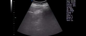
- Ultrasound signs of PI (continued enlargement of the gland, blurred contours and the appearance of fluid in the tissue).
To monitor PI it is recommended:
- dynamic study of clinical and laboratory parameters;
- at least 2 ultrasound scans in the 2nd week of the disease;
- at the end of the 2nd week, CT scan of the pancreas.
Possible outcomes by the end of phase IB:
- resorption for up to 4 weeks;
- aseptic sequestration of pancreatic necrosis with the formation of a pseudocyst: stabilization of the size of the PI with normalization of health and subsidence of SIRS against the background of hyperamylasemia;
- septic sequestration (purulent complications).
Protocol for the diagnosis of purulent complications in phase II of the disease in the phase of septic sequestration.
Infection of the site of destruction occurs at the end of the 2nd – beginning of the 3rd week.
With late admission, inadequate treatment or too early surgery, infection may bypass the period of aseptic destruction (“crossover phases”).
Infected pancreatic necrosis:
- limited – pancreatic abscess (PA);
- not delimited – purulent-necrotic parapancreatitis (NPP).
Verification of purulent complications:
- progression of clinical and laboratory indicators of acute inflammation in the 3rd week;
- on MSCT, MRI, ultrasound, growth of devitalized tissue and/or gas bubbles;
- positive bacterioscopy and bacterial culture of aspirated samples during fine-needle puncture.
Examination by a surgeon during the first hour of hospitalization.
3. Treatment.
Each phase of the disease corresponds to a specific clinical and morphological form of AP; it is most advisable to consider treatment tactics for AP in the corresponding phases of the disease.
3.1 Conservative treatment.
Treatment of acute pancreatitis in phase IA of the disease Intensive conservative therapy is recommended.
Laparotomy is indicated only in the event of the development of surgical complications that cannot be eliminated by minimally invasive technologies.
I. Treatment of mild AP.
Hospitalization in the surgical department.
Basic treatment complex:
- hunger;
- probing and aspiration of gastric contents;
- local hypothermia (cold on the stomach);
- analgesics;
- antispasmodics;
- infusion therapy up to 40 ml/1 kg of body weight with 24-48-hour forced diuresis.
Basic therapy is enhanced with inhibitors of pancreatic secretion.
If there is no effect from the therapy within 6 hours and at least one more sign of the express assessment scale, moderate (severe) pancreatitis is stated.
In case of moderate (severe) pancreatitis, treatment is carried out in the intensive care unit according to protocols III, IV.
II. Intensive therapy of moderate acute pancreatitis.
Hospitalization in the ICU; in the absence of organ failure and progression within 24 hours, transfer to the surgical department is possible.
The main type of treatment is conservative therapy.
The basic treatment complex is supplemented by a specialized treatment complex.
Specialized treatment:
- pancreatic secretion inhibitors, optimally the first three days;
- active rheological therapy;
- infusion therapy of at least 40 ml/1 kg of body weight with forced diuresis in case of organ dysfunction (in the absence of contraindications);
- antioxidant and antihypoxic therapy;
- evacuation of toxic exudates according to indications;
- for enzymatic peritonitis - sanitary laparoscopy, percutaneous drainage under ultrasound guidance or laparocentesis is allowed.
Prophylactic antibiotics are not recommended.
III. Intensive therapy of severe acute pancreatitis.
Hospitalization in the ICU.
The main type of treatment is intensive therapy.
The basic treatment complex is supplemented by a specialized treatment complex.
Recommended:
- extracorporeal detoxification methods: plasmapheresis; hemofiltration;
- nasogastric tube for decompression;
- if possible, nasogastrointestinal intubation for early enteral support;
- correction of hypovolemic disorders;
- epidural block;
- disaggregant antithrombotic therapy.
Antibiotics are not recommended for prophylactic purposes in the first three days of the disease.
Phase IB treatment, i.e. peripancreatic infiltrate.
In most patients, treatment is conservative.
Laparotomy in the 2nd week of AP only in case of surgical complications that cannot be eliminated by minimally invasive technologies: destructive cholecystitis, gastrointestinal bleeding, acute intestinal obstruction, etc.
Composition of the treatment complex:
- continuation of basic infusion-transfusion therapy;
- therapeutic nutrition table No. 5 (moderate AP);
- nutritional support: oral, enteral or parenteral (severe AP);
- systemic antibiotic prophylaxis (cephalosporins of III-IV generations or fluoroquinolones of II-III generations in combination with metronidazole, reserve drugs - carbapenems);
- immunotherapy (correction of cellular and humoral immunity).
Late (II) phase (sequestration)
Treatment of AP in the phase of aseptic sequestration, i.e. treatment of pancreatic pseudocyst.
It is not recommended to operate on small pseudocysts – less than 5 cm; dynamic observation by a surgeon.
Large pseudocysts - more than 5 cm - are operated on routinely in the absence of complications.
The operation of choice for an immature pseudocyst (less than 6 months) is external drainage.
A mature pseudocyst (more than 6 months) is subject to surgical treatment as planned.
Complications of pancreatic pseudocyst:
- infection;
- bleeding into the cyst cavity;
- perforation with a breakthrough into the abdominal cavity with the development of peritonitis;
- compression of neighboring organs with the development of obstructive jaundice, gastric stenosis, intestinal obstruction, etc.
Deadlines
The duration of inpatient treatment depends on the condition of the person in which he was hospitalized and the accurate implementation of medical prescriptions. Symptoms of mild pancreatitis most often resolve within a few days with intensive drug infusions. Treatment of exacerbations in patients with chronic inflammation of the gland requires much longer hospitalization, especially during surgery.
Acute form of the disease
For a primary attack of moderate pancreatitis, a course of therapy of about 2-3 weeks is required. During this time, in most clinical cases, it is possible to completely improve the pancreas. However, 6 months after discharge from the hospital, treatment must be repeated so that the disease does not become chronic.
Chronic stage
People suffering from a long-term form of pancreatitis need scheduled hospitalization every six months to prevent exacerbations. This is facilitated by medicinal healing of the pancreas and the entire body. The duration of the course of treatment can range from 10 to 21 days.
[morkovin_vg video=”G1Gsr6ggvdM;jwToPsco0gU;Aef8GoCmD5Y”]
Prevention and follow-up
- At the outpatient stage, D II and D III groups.
- Scheduled examinations by a therapist and gastroenterologist 1-4 times a year, depending on the frequency of exacerbations.
- If indicated, consult a surgeon.
- If blood sugar increases, consult an endocrinologist.
- Clinical blood and urine analysis, blood amylase and urine diastase, blood and urine sugar.
- Ultrasound of the liver, gallbladder, pancreas,
- Cholecystography according to indications.
- Preventive courses of drug treatment 2-4 times a year, according to indications - physiotherapy, exercise therapy.
- Diet correction, rational employment, selection for sanatorium treatment.
- If there are signs of disability - MSEC.
- Indications for sanatorium-resort treatment are recurrent chronic pancreatitis with rare relapses without a tendency to frequent exacerbations.
- Treatment at balneological resorts: Zheleznovodsk, Essentuki, Morshin, Truskavets, Izhevsk Mineral Waters, etc.
- Climatotherapy: air baths, swimming in open water (stable remission).
- Medical nutrition – table No. 5.
- Non-carbonated, low- and medium-mineralized mineral waters, with bicarbonate and sulfate ions, alkaline waters.
Procedures:
- carbon dioxide-hydrogen sulfide baths;
- carbon dioxide-radon sulfide;
- pearl (every other day);
- heat therapy;
- mud and ozokerite applications;
- electrophoresis of dirt;
- sinusoidal-modeled currents;
- SMV therapy;
- DMV therapy;
- diadynamic currents.
Therapeutic physical education according to a gentle tonic regimen with morning hygienic exercises, sedentary games, dosed walking.
Number of people who read this article: 4,134
Treatment of pancreatic necrosis is carried out in a hospital
In case of sterile necrosis, doctors prescribe conservative therapy, but in case of purulent fusion, they must perform an operation - removal of foci of pancreatic necrosis using an open method or laparoscopically. Genetic studies indicate that people with Toll-like receptor-4 polymorphisms have an increased risk of infection of the pancreas when it becomes inflamed. TLR-4 is located both in the intestine and in the pancreas and liver. They react to bacterial molecules. In people with increased sensitivity of these receptors, pancreatitis occurs with pancreatic necrosis.
In the postoperative period, complex therapy is indicated:
- Antibiotic therapy;
- Medical nutrition (table number 5 for moderate AP);
- Immunotherapy;
- Antioxidants;
- Prevention of bacterial overgrowth syndrome;
- Normalization of bile outflow.


