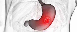
Etiology and pathogenesis of eventration
Eventration appears 6-10 days after surgery, during a period of slow healing processes. It is believed that this complication often develops when sutures are placed incorrectly. For a defect to appear, a combination of several factors is necessary.
The main causes of eventration:
- suppuration of the suture;
- diabetes;
- non-compliance with diet;
- inflammatory processes in the wound;
- increased physical activity after surgery;
- improperly sutured wound;
- packing the wound through the abdominal cavity;
- atrophic processes in the abdominal wall - occur in malnourished patients.
When exposed to several causes, the operated area is weakened, which leads to improper restoration of connective tissue. Pressure on the affected area from the inside poses the greatest danger to the patient's health.
The divergence of the edges of the wound is facilitated by such circumstances as cough, bloating, vomiting, intestinal paresis - impaired motor function (weakening of movements or their complete cessation).
In some cases, eventration of the small intestine occurs as a result of an incision that is too wide when forming a stoma - an artificial exit of a loop to the outside to drain feces. This need arises in case of obstruction, oncology, severe forms of ulcerative colitis. In adult patients, eventration during a small intestinal stoma is caused by a violation of the therapeutic and protective regime (heavy lifting). In this case, emergency surgery is required.
Reasons for the development of pathology
The occurrence of eventration is due to the following reasons:
- metabolic disorders;
- deficiency of blood clotting factor 13;
- hypovitaminosis;
- anemia,
- obesity;
- slow healing of wounds;
- vomiting and cough;
- excessively active movements.
There are also surgical reasons that contribute to the development of such a phenomenon as eventration:
- incorrect technique for applying surgical sutures to the abdominal cavity;
- drainage through the wound;
- removal of sutures before the deadline;
- purulent processes in the wound.
In the vast majority of cases, eventration develops 6-10 days after surgery. At this moment the seams begin to come apart.
Classification of pathology
The classification of visceral prolapse is especially important, since a clear division of the forms of postoperative conditions makes it possible to clarify the indications for repeated surgical interventions. The most popular classification is E.S. Baimysheva 1989.
According to the degree of organ prolapse, eventration is:
- Open (external). Organs fall out through the wound opening onto the surface of the body.
- Internal (interpleural). The viscera penetrate the pleural cavity through the diaphragm.
- Subcutaneous. More often it occurs after laparotomy (an incision in the abdominal wall) as a result of suture divergence. All layers of the abdominal wall, except the skin, are separated.
Dehiscence of the wound edges can be complete or partial. Based on the nature of infection, postoperative prolapse of organs into a clean or purulent wound, as well as peritonitis outside the affected area, are distinguished.
The difference between a hernia and an eventration is the presence of a hernial sac lined with peritoneum. In organ prolapse, the internal organs come out or under the skin.
The first observation of internal organ prolapse through the vagina was described in 1864 by Hyernaux [3]. McGregor in 1907 described the observation of spontaneous vaginal rupture in a 63-year-old woman after lifting “half a ton of coal” [8]. Prolapse of internal organs through the vaginal stump after laparoscopic hysterectomy is quite rare [1, 7, 10-13]. At the beginning of the 20th century, P. Ramirez et al. [9] reported 59 cases of eventration through the vaginal stump after hysterectomy from 1900 to 2001 with an incidence of 0.3%.
H. Hur et al. [2] reported failure of the vaginal stump in 4.9% of cases after laparoscopic hysterectomy, in 0.29% after vaginal hysterectomy and in 0.12% of cases after laparotomic hysterectomy on average 11 weeks after surgery. In earlier publications by P. Iaco et al. [4] stated the incidence of internal organ prolapse at 0.79, 0.25 and 0.26% of observations, respectively. In young patients, prolapse of internal organs has been described in the first 6 months after surgery. In old age, prolapse of internal organs through the vaginal stump was observed at a later date, in particular several years after surgery, and is associated with weakness of the pelvic floor muscles [4-6].
Although eventration of various abdominal organs through the vaginal stump is described in the literature, we have not found a single mention of infringement of an eventrated organ.
Here is an observation.
Patient G.,
46 years old, hospitalized in the surgical department of the Reutov City Clinical Hospital on 10/09/11 as an emergency on the 1st day from the onset of the disease with complaints of constant aching pain in the hypogastric region, dry mouth, bloating.
Considers himself sick during the last 24 hours, when at about 14:00 08.10 pain appeared in the hypogastrium after coitus. She did not treat herself. Subsequently, the pain intensified and became permanent. Hyperthermia reached 38 °C. Due to increased pain, she turned to the Reutov Central City Hospital.
Among the past diseases, he notes childhood infections. Operations: appendectomy in 1985, hysterectomy for endometrial hyperplasia, uterine fibroids in June 2010.
At the time of examination, the condition was of moderate severity. Consciousness is clear. The patient has a normal build and normal nutrition. The skin and visible mucous membranes are of normal color. Body temperature 38.3 °C. NPV 16 per minute. Auscultation: vesicular breathing, no wheezing. Heart sounds are clear, rhythmic, pure, no pathological noises are heard. Heart rate 78 per minute, blood pressure 130/80 mm Hg. The tongue is dry, covered with a white coating. The abdomen is of regular shape, moderately swollen, in the right iliac region there is a postoperative keloid scar measuring 12x1 cm without signs of inflammation. On palpation, the abdomen is soft, sharply painful in the hypogastric region, where protective muscle tension is detected. No infiltrative or tumor-like formations were detected in the abdominal cavity; the Shchetkin-Blumberg sign was positive in the hypogastric region. Peristaltic sounds are sluggish, no pathological bowel sounds are heard. Tapping on the lumbar region is painless. Physiological functions are not impaired.
Per rectum: sphincter tone is preserved, tumor-like formations are not detected at the height of the finger. On palpation along the anterior wall of the rectum, sharp pain is detected. Per vagina: the presence of a fatty pendant and a section of the colon in the vaginal lumen was revealed.
Blood leukocytes 12.2·109/l.
09.10 - surgery for urgent indications: elimination of eventration. Laparoscopic suturing of the vaginal stump, sanitation, drainage of the abdominal cavity.
Carboxyperitoneum was applied through a Veress needle to an intra-abdominal pressure of 12 mm Hg. A video camera and manipulators (1×5, 1×10 and 1×10 mm) are introduced through typical points. An examination of the liver, gallbladder, and spleen revealed no pathological changes. In the abdominal cavity there is up to 100 ml of cloudy serous effusion, mainly in the pelvic cavity. The effusion was aspirated. From the vaginal stump, a strangulated portion of the sigmoid colon with a fat suspension was inserted into the abdominal cavity (see figure).
Figure 1. Intraoperative photographs of patient G. a - parietal entrapment of the fatty suspension of the sigmoid colon.
Figure 1. Intraoperative photographs of patient G. b - incompetent vaginal stump.
Figure 1. Intraoperative photographs of patient G. in - intracorporeal suture of the vaginal stump. The latter with injected vessels. During instrumental examination, the wall of the sigmoid colon is pinched parietally and is viable. The vaginal defect measuring 3x4 cm was sutured with two single interrupted 2/0 Vicryl sutures. A drainage is installed in the pelvic cavity to the suturing site. The abdominal cavity was sanitized with 500 ml of antiseptic and drained. Desufflation was performed and the wounds were sutured.
Postoperative course without complications. The patient was discharged 7 days after surgery.
According to various authors, the number of eventrations is progressively increasing with the introduction of video endoscopic technologies into practice, so, according to various sources, their number after laparoscopic and robot-assisted hysterectomy reaches 5%. H. Hur et al. [2] provide data from a survey of 7039 patients after hysterectomy. In 8 out of 10 cases, failure of the vaginal stump occurred after laparoscopic hysterectomy, which is 2%. All 8 patients were young (average age 39 years), and the technique of vaginal suturing was no different from the traditional one. The authors believe that the access and technique of suturing the vaginal stump do not affect the incidence of vaginal stump failure [2, 4]. P. Iaco et al. [4] compared 1440 patients who underwent hysterectomy with suturing of the vaginal stump and 2330 without suturing. There was no significant difference in the incidence of internal organ prolapse.
It is necessary to make a reservation that when a patient is admitted with a clinical picture of nonspecific abdominal pain in the absence of a picture of intestinal obstruction on an x-ray and unexpressed changes in laboratory tests, the differential diagnosis of abdominal pathological changes is difficult. Acute abdominal pain in women can be a manifestation of various pathological changes in internal organs, including acute gynecological diseases.
In our observation, the cause of pelvioperitonitis before laparoscopy remained completely unclear.
Panhysterectomy is currently completed with a hardware or manual suture of the vagina. In this case, patients are recommended to have “sexual rest” for 2-3 months. In the patient we observed, the postoperative period after hysterectomy, occurring with symptoms of prolonged colporrhagia, and the early onset of sexual activity contributed to the formation of incompetent vaginal stump with subsequent prolapse of the sigmoid colon and Richter's strangulation. The use of laparoscopy made it possible to establish the correct diagnosis and carry out timely surgical treatment.
Clinical picture
Partial and subcutaneous eventration develop gradually. Most patients experience intestinal obstruction, symptoms of intoxication and peritonitis. Clinical manifestations:
- pain in the wound area;
- temperature increase;
- deterioration of general condition;
- Wetting of the bandage - occurs as a result of the release of pus or fluid accumulating in the abdominal cavity.
The clinical picture of subcutaneous eventration is mild – the patient’s general condition does not worsen and no pain occurs.
Diagnostic methods
The diagnostic process in this case does not cause any difficulties. The very first symptom of eventration is the blotting of a dry gauze sticker with a small amount of serous and bloody discharge. This suggests that the deep layers have ruptured, and only the skin is a barrier to intestinal prolapse. A sign of the onset of eventration is the development of emphysema in the wound area and pain in it.
Intestinal eventration does not imply absolute prolapse of the organ. Fibrin, which allows the intestine and parietal peritoneum to stick together, prevents the organ from falling out. When such a situation develops, almost all patients talk about stopped or difficult removal of gases, the appearance or intensification of the feeling of bloating. A significant proportion of patients exhibit signs of partial or complete intestinal obstruction and develop symptoms of peritonitis or intoxication. The final conclusion about the development of eventration can only be made during dressing.
Diagnostics
When the wound is completely dehisced, the diagnosis is made quickly and accurately. Subcutaneous eventration is more difficult to determine. Often this type of disease is not detected in a timely manner. Probing the wound “blindly” in this case is unacceptable, since there is a high probability of damaging the underlying intestine.
The complication can be identified at an early stage by removing several stitches and examining the wound. Lateral radiography with the patient in the supine position also helps determine the complication. Against the background of a clearly visible line of the aponeurosis (wide tendon plate), in the presence of pathology, a light spot is visible.
During subcutaneous eventration, it is recommended to perform an ultrasound of the anterior abdominal wall, which allows one to determine its thinning in the scar area. When conducting this study, the difference between eventration and hernia becomes apparent.
Therapy methods
Eventration is an indication for emergency surgery. If the wound has not festered and tissue inflammation has not occurred, the prolapsed entrails are treated with an antibiotic solution, inserted into the abdominal cavity, and the wound is sutured with mattress sutures. Dacron, nylon thread or thick silk are used as suture material. Sutures are allowed to be removed no earlier than 12-14 days.
If the wound is suppurated or inflamed, after carefully repositioning the prolapsed organs, tampons with fish oil or vaseline oil are placed in the wound, and then an aseptic bandage is applied. In some cases, a plaster splint is placed on the anterior abdominal wall.
If eventration occurs after a penetrating wound to the abdomen, damage to internal organs must be assumed and an inspection of the abdominal cavity is performed. The tightness of the wall of the intestinal loop and its blood supply are checked. If they are not broken or covered with fibrin deposits (a protein produced in the liver), the intestinal loop is washed with an antiseptic solution and placed back into the abdominal cavity. In the treatment of eventration of the omentum, its resection (cutting out) is performed.
Internal eventration is an indication for urgent excision or reduction of organs and suturing of the diaphragm.
Features of the bandage during eventration:
- if organs have fallen out of the wound, you cannot set them back when providing first aid; you must apply an antiseptic cloth and moisten it regularly;
- for intestinal prolapse, a donut bandage is most effective;
- the main task in case of injury is to stop the bleeding, which will require creating pressure on the affected area;
- the skin around the site of organ prolapse is treated with brilliant green or iodine.
The speed of measures taken and the qualifications of doctors are of great importance. After surgery, the patient must strictly follow all instructions.
Treatment of eventration
Eventration therapy can be carried out both by conservative methods and through surgical intervention. When a patient experiences subcutaneous eventration and the patient’s general condition does not worsen, doctors resort to conservative treatment methods. In this case, bed rest and application of a bandage to the anterior abdominal cavity for 15-20 days are recommended. Operations on the anterior abdominal wall for reconstruction purposes are carried out after 3-5 months. Conservative treatment may also be indicated for partial eventration. The essence of treatment in this case is to eliminate the purulent-necrotic process in the surgical area and prepare the wound for the application of secondary sutures, as well as to take measures to eliminate possible relapses of the condition.
In case of ineffectiveness of conservative treatment methods, as well as eventration of degrees 3 and 4, surgical intervention is recommended. The operation is performed 1-2 hours after preparation. During the manipulation, old sutures are removed, the edges of the wound are sparingly removed, and sutures are applied through all layers. These sutures are eliminated between 18 and 22 days after the eventration has been sutured.
In conclusion, I would like to note that eventration itself cannot be the cause of the patient’s death. However, against the background of other aggravating circumstances, the course of the postoperative period significantly worsens. As is known, after surgery, the patient’s immunity level decreases due to eventration. For this reason, he is prescribed immunostimulating drugs and immunostimulating methods such as blood transfusions and anabolic hormones.
Preventive measures
Among the areas of preventive work during eventration are the impact on local and general factors, as well as the fight against increased intra-abdominal pressure. When local factors are eliminated, prevention is aimed at preventing wound suppuration, treating peritonitis and carefully suturing the damaged area.
To reduce the number of infectious complications, antibiotic therapy is used. The drug must be in the blood until the start of the operation. To reduce the risk of wound suppuration, additional treatment of the surgical field with antiseptic solutions is carried out and various protective films are used. Antistaphylococcal plasma, gamma globulin and toxoid in combination with antifagin are also used.
Prevention of eventration, which consists of influencing general factors, involves treating the underlying disease and eliminating homeostasis disorders (anemia, hypovitaminosis). Particular attention is paid to detoxification therapy. Since organ prolapse can occur as a result of delayed wound healing, it is recommended to use substances that regulate regeneration processes and eliminate inflammation.