Clinical picture
The clinical picture depends on the nature of organ damage, the intensity of bleeding into the abdominal cavity and the amount of blood loss.
In acutely occurring G., a picture of acute anemia is observed: pallor of the skin and visible mucous membranes, thirst, cold sweat, adynamia, darkening of the eyes, dizziness, fainting, and there may be motor agitation; Due to painful irritation of the dome of the diaphragm that has gushed out blood, the patient tends to take a sitting position (the vanka-stand-up symptom), which reduces the pain. The pulse is frequent (up to 120-140 beats per minute), weak filling, blood pressure decreases. The abdomen participates in breathing to a limited extent, is soft and moderately painful on palpation, and there are symptoms of peritoneal irritation. In the sloping parts of the abdomen there is dullness of percussion sound, percussion is painful, bowel sounds cannot be heard. During a digital examination of the rectum, a protrusion of its wall may be detected; during a vaginal examination, the fornix is flattened and painful; the posterior fornix may bulge. When examining peripheral blood, there is a decrease in hemoglobin, the number of red blood cells, and the hematocrit value. In the initial stages of G.'s development, changes in peripheral blood are not pronounced. The more time has passed since the onset of bleeding, the more clearly the clinical symptoms of G. are revealed. When bleeding into the abdominal cavity from small vessels in the postoperative period, G. develops slowly, symptoms from the abdomen are often erased and difficult to detect against the background of changes associated with surgical trauma. With G., a frenicus symptom may be observed - pain in the area of the shoulder girdle and shoulder blade, often on the left, due to irritation of the endings of the sensory nerves of the diaphragm by blood pouring into the abdominal cavity (see Frenicus symptom).
Hemoperitoneum
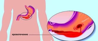
Hemoperitoneum (laparohemorrhage, bleeding into the abdominal cavity, intra-abdominal bleeding) is a common condition in thoracoabdominal surgery, gastroenterology, gynecology, traumatology, and disaster medicine. With hemoperitoneum, there is an outflow and accumulation of free fluid (blood) between the visceral and parietal layers of the peritoneum. Suspicion of hemoperitoneum requires emergency hospitalization and surgical assistance. The death of patients with intra-abdominal internal bleeding most often occurs from traumatic shock and acute blood loss.
Diagnosis
The diagnosis with a clear wedge picture is beyond doubt. When the wedge is erased, the phenomena of G. have diagnostic value: laparocentesis (see) with the introduction of a groping catheter and peritoneoscopy (see), as well as puncture of the posterior vaginal vault. The introduction of a “groping” catheter and examination of the blood obtained from the abdominal cavity make it possible to establish the nature of the bleeding - whether it continues or has stopped, this is judged by the Rouvilois-Gregoire test (see Hemothorax). Data on the specific gravity of blood are of great importance in determining the amount of blood loss. Surgical tactics in each specific case depend on these diagnostic techniques.
Patients with suspected G., before establishing a final diagnosis, require careful dynamic monitoring with measurement of pulse rate and blood pressure every 1-2 hours, determination of the amount of hemoglobin and the value of peripheral blood hematocrit. During the diagnostic process, painkillers and narcotics that mask the symptoms of an acute abdomen are contraindicated (see).
G. must be differentiated from retroperitoneal hematoma and hematoma of the anterior abdominal wall, in which there are no symptoms of free fluid in the abdominal cavity.
X-ray diagnosis of hemoperitoneum
aims to establish the presence of free fluid in the abdominal cavity. The main methods of investigation for G. are fluoroscopy and radiography of the abdominal cavity. Pneumoperitoneum (see), and even more so angiography of the vessels of the abdominal cavity are permissible only if it is necessary to preoperatively clarify the nature of damage to the abdominal or pelvic organs or to first locate a bleeding vessel. These studies can only be carried out if the patient’s condition is satisfactory, with appropriate equipment and specially trained personnel. In case of traumatic origin of G., it is necessary to examine other damaged organs. All rentgenol examinations should be carried out with maximum sparing of the patient, in a supine position, and when examining the pelvic area - in a semi-sitting position.
When fluoroscopy of the abdominal cavity in patients in the first hours after the development of gastrointestinal tract, there is a restriction in the respiratory excursions of the diaphragm, and at a later date, signs of intestinal paresis are revealed - swollen intestinal loops with levels of gas and liquid in them.
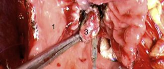
Diagrams of radiographs of the pelvic area with hemoperitoneum: a - with a small amount (about 150 ml) of blood; b - when the amount of blood is about 300 ml; 1 - pubic symphysis; 2 - sacrum; 3 - light shadow of the parietal fatty tissue of the pelvis; 4 - accumulation of blood.
On a survey radiograph, it is necessary to obtain an image of the subdiaphragmatic and lateral areas of the abdomen, as well as the pelvic area. Small amounts of blood, due to gravity, accumulate in sloping areas of the abdominal cavity and are detected radiographically in the form of characteristic areas of darkening (Fig., a).
A larger amount of fluid on plain radiographs of the abdominal cavity is determined in the form of wide bands of darkening in the area of the lateral sections with internal scalloped contours (Fig., b), as well as in the form of triangular, oval or polygonal shadows with vague contours between the intestinal loops swollen with gas (S. A. Reinberg). Lateroscopy in these cases reveals a symptom of intestinal floating (G. A. Zedgenidze, L. D. Lindenbraten, etc.). Radiography of the pelvic area is of great importance (for diagnosing small amounts of fluid). With peritoneography (see), it is possible to even more accurately determine the location and paths of distribution of small volumes of free fluid, the edges, being contrasted, evenly cover the surface of the abdominal organs, allowing them to obtain a clear X-ray image. Diagnostic pneumoperitoneum (see) with the introduction of 200-300 ml of gas also greatly facilitates the identification of a small amount of free fluid in the abdominal cavity due to the formation of a horizontal level at the border between liquid and gas.
G. X-ray must be differentiated from a retroperitoneal hematoma; the edges on a radiograph are usually revealed by the expansion of the shadow and the disappearance of a clear contour of the lumbar muscle on one side.
Causes of hemoperitoneum
Hemoperitoneum can complicate a wide range of conditions: closed abdominal injuries, thoracoabdominal injuries, pathological processes in the abdominal cavity and retroperitoneal space, surgical interventions, gynecological diseases, etc. Hemoperitoneum develops as a result of a violation of the integrity of the vessels of the abdominal cavity and can be of traumatic and non-traumatic origin.
Traumatic hemoperitoneum occurs with blunt blunt trauma to the abdomen (blows, falls, road accidents, compression), penetrating (gunshot, knife) wounds of the abdomen, intraoperative trauma. In this case, parenchymal organs are most often damaged - the liver, spleen, pancreas, as well as vessels passing through the ligaments, folds of the peritoneum, intestinal mesenteries, and omentum. Damage to internal organs from bone fragments can occur when the lower ribs are fractured.
Non-traumatic hemoperitoneum occurs in ectopic pregnancy, ovarian apoplexy, rupture of aortic aneurysm, liver hemangioma, diseases leading to decreased blood clotting (obstructive jaundice, malaria, blood diseases, hemorrhagic diathesis), long-term therapy with fibrinolytics and anticoagulants. In surgical practice, hemoperitoneum can develop as a result of organ damage during invasive diagnostics (angiography, puncture biopsy), cutting or slipping of surgical ligatures after gastric resection, appendectomy, hernia repair, hemicolectomy, cholecystectomy, supravaginal amputation of the uterus, nephrectomy, splenectomy, liver resection and etc.
Forecast
The prognosis for G. is always serious; it depends on the nature of the injury or disease that caused the development of G., the amount of blood loss and the period that elapsed from the beginning of G.’s development to the operation.
Undiagnosed G. often becomes infected and leads to the development of diffuse or limited peritonitis (see).
See also Bleeding.
Bibliography:
Zedgenidze G. A. and Lindenbrate N L. D. Emergency X-ray diagnostics, L., 1957; Littmann I. Abdominal surgery, trans. from German, Budapest, 1970; Emergency surgery, ed. N. I. Blinova and B. M. Khromova, L., 1970; Experience of Soviet medicine in the Great Patriotic War of 1941-1945, vol. 12, M., 1949; P and p-to about A. S. X-ray diagnosis of early complications after gastrectomy # M., 1958, bibliogr.; F g iman n- D ah 1 J. Roentgen examination in acute abdominal diseases, Springfield, 1951
G. A. Pokrovsky; L. M. Freidin (rent.).
Diagnostics
Due to the non-obviousness of the signs of hemobartonellosis, veterinarians often have no idea about the main cause of the dog’s illness, and therefore treat the wrong disease. Although, if you suspect the presence of an infection in time, it is quite possible to detect it. The most reliable diagnostic methods for identifying bortanella in a dog’s body are:
- examination of cells obtained by biopsy;
- use of serological tests;
- use of polymerase chain reaction;
- growing an infectious agent on a nutrient medium.
Read Etiology of actinomycosis in dogs: signs, therapy and prevention
For greater accuracy of the result, it is advisable to use several of the above methods at once, since none of them is 100% accurate. Bartonella tends to “hide” in red blood cells - this makes their detection to a certain extent difficult.
First aid
If the hemorrhage does not stop for a long time, then the dog’s body temperature may rise significantly (to critical levels), and its general condition worsens. In this case, you should not hesitate and it is better to provide emergency assistance yourself. It is recommended to do the following:
- Apply an ice or cold compress to the area of injury: ice placed in a bag, a cloth moistened with cold water, or a heating pad filled with it. Such an event will help stop bleeding and reduce pain, but it should be used correctly. To prevent ice from damaging your skin, you should first wrap it in cloth. The compress can be kept on one area for no longer than 15 minutes, which will eliminate the possibility of frostbite to the tissue. After 2-3 hours, it is recommended to repeat the procedure.
- The damaged area can be treated with iodine solution - you can apply a mesh, which has an anti-inflammatory effect.
- A tight bandage is applied to the hematoma. It is best to use bandages for this. But if the dressing is done in the field, then you can take any sterile material for these purposes, a clean handkerchief or napkin will do.
This is interesting: Gamavit can be used by humans
If the subcutaneous hemorrhage reaches a large size, then a paraffin compress can be applied. In addition, exposure to heat helps speed up the resorption process: warming up under a special lamp, using warming ointments. But thermal procedures should be carried out after the swelling subsides, no earlier than a day after the hematoma appears.
Watch a video about ear hematoma in dogs:
Treatment of hemobartonellosis in dogs
If an infection is detected, the veterinarian prescribes treatment and constantly monitors the animal’s condition. The dog is usually treated at home; placement in a veterinary hospital is not necessary.
There are no special medications that can cure a dog of bartonellosis. But treatment with antibiotics is quite effective. Most often they resort to tetracycline injections - they must be done at intervals of 8 hours. The use of azithromycin, rifampin, doxycycline, and enrofloxacin also gives positive results. All of the above medications destroy the cell membrane of batonella, thereby contributing to its death. For the dog to fully recover, the course of antibiotic therapy must last at least three weeks.
In parallel with antibiotics, medications are used to alleviate the general condition of the animal. They are selected depending on the symptoms. For example, if the disease is accompanied by diathesis, the animal’s skin is treated with ointments that relieve inflammation and itching and promote wound healing.
It is recommended to give your dog B vitamins and preparations containing iron that improve hematopoiesis.
You should pay attention to the animal's diet. It is advisable to give your dog more meat and not replace it with dry dog food. Liver is especially recommended as it improves blood quality.
It is possible to determine that an animal is healthy only through control tests. After recovery, the dog acquires immunity from bartonellosis for life.