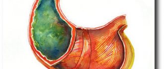
The stomach and duodenum are part of the digestive tract, where food accumulates and is partially digested, gradually moving to the next sections of the intestine. The stomach is a kind of expansion of the esophageal tube, shaped like a pouch. The organ has inlet and outlet openings, and towards the bottom it passes into the tubular duodenum. The stomach is separated from it by a special valve - the sphincter, which opens only when the next portion of food enters the stomach.
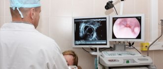
The most relevant study of this system in the human body is considered to be fibrogastroduodenoscopy, during which the doctor, using a special endoscope, directly examines the mucous membranes of the esophagus, stomach and duodenum, and can also take tissue samples for biopsy. However, in cases where the results obtained from FGDS or, for example, from fluoroscopy with barium contrast are not enough to establish a diagnosis and determine treatment tactics, doctors turn to magnetic resonance imaging.
Cases where the procedure is necessary
The urgency of the procedure appears when:
- the specialist assumes that the human stomach or esophagus is affected by a malignant tumor, or a benign formation has arisen;
- the patient is diagnosed with cancer of the stomach or esophagus;
- the body became infected with parasites, which significantly undermined human health;
- there is a need to diagnose a stomach ulcer, chronic gastritis or hernia;
- it is necessary to determine the specific area of the location of metastases in the stomach;
- there are suspicions of the development of a stomach ulcer, which entails complications in the form of a through defect in the wall of a hollow organ or a pathological process in the tissues.
List of indications and contraindications
A procedure associated with a detailed examination of the stomach, under special circumstances, is indicated for people who are suspected of having: formations of a benign or malignant nature, metastases that have spread from other organs, diseases of the gastrointestinal tract, for example, diaphragmatic hernia, gastritis or stomach ulcers. The list continues with the presence of a complex form of helminthic infestation, abnormal structure of an abdominal organ, perforation or malignancy of a peptic ulcer.
The study can also monitor the effectiveness of treatment methods developed to combat stomach cancer. The procedure, despite its relative safety and painlessness, is not carried out in every case. Sometimes the health condition of the subject is a serious reason for refusing to undergo a radiation procedure.
Gastric biopsy - what is it?
The category of contraindications includes:
- Lactation period (breastfeeding) and gestation (pregnancy).
- Severe form of liver failure.
- Presence of a previously implanted heart valve.
- An image on the patient’s body of decorative tattoos made with dyes containing various metallic inclusions.
- The presence in the human body of ferromagnetic metals in the form of bullets, fragmentation elements, vascular clips, pacemakers, bone fixators, plates and wires inserted in fractures.
In addition to the main prohibitions, there are also relative contraindications: heart failure with complications, claustrophobia (pathological fear of closed spaces), mental abnormalities accompanied by uncontrolled movements of the patient, extremely serious condition of the patient.
For people who exhibit such ailments, MRI is prescribed only when absolutely necessary to further minimize dangerous consequences. Medical personnel take the necessary measures to reduce various risks to the patient’s life to zero.
What can a tomograph image show?
On the images that are handed over to the patient who has undergone MRI, the specialist is able to determine:
- localization of pathology and features of its formation;
- metastases, their nature and size;
- the place of development of the inflammatory process of the gastrointestinal tract caused by an ulcer or gastritis;
- the presence of bleeding of the gastric walls;
- the cause of the development of the pathological process.
In addition, the doctor evaluates the condition of the lymph nodes that surround the organs of the digestive system and determines whether there are foreign objects in the human gastrointestinal tract.
The procedure makes it possible to diagnose a large number of pathologies, which can be identified at an early stage of development and treatment can be started in a timely manner.
What does it show?
An MRI scan of the stomach is done if there are accumulated gases in the abdominal cavity, which may indicate malignant tumors and ulcers. MRI shows possible malignant formations, the condition of the abdominal organs that are located close to the stomach. An MRI is done to determine the condition of the stomach walls and exclude or confirm pathological changes in the organ.
MRI allows you to diagnose malignant tumors, possible metastases, size and location of the tumor. MRI makes it possible to predict the development of the tumor and its relationship with other internal organs.
What to expect from the procedure and how to prepare?
The examination is absolutely painless. You should take off all items made of metal in advance and temporarily part with your phone, keys, credit card, etc.
The patient is placed in a tomograph tunnel, the diameter of which is 70-80 cm. For the patient’s comfort during the procedure, the tunnel is equipped with ventilation and lighting. During the examination it is necessary to remain in a stationary position. The result is a high-quality image of three projections of the stomach.
In order for an MRI of the esophagus and stomach to be painless, you should devote a little time to preparation, which involves:
- abstaining from eating 5-6 hours before the study;
- selection of clothing made from natural fabrics;
- taking sedatives when undergoing an MRI for a child under 5 years of age:
- taking activated carbon several hours before the procedure in case of increased gas formation;
- following a low-carbohydrate diet for 3 days before the study (as prescribed by the doctor).
Several hours after the tomography, you can return to your normal life.
When is an MRI of the stomach prescribed?
Magnetic resonance imaging of the gastrointestinal tract can be performed at the patient’s request, for example, if he wants to make sure that everything is fine with him and there are no tumor processes.
Indications for MRI are:
- abdominal pain of a cutting, sharp, dull, spastic or aching nature, especially if they appear with a certain frequency;
- if after an ultrasound or CT scan there is a suspicion that there is a tumor in the stomach;
- assessment of the stage of stomach disease;
- preparation for surgical intervention on the abdominal organs;
- congenital anomalies of the stomach;
- internal bleeding;
- increased gas formation;
- nausea and vomiting due to an unknown reason, change in the color of urine and feces;
- clarification of ultrasound and CT results;
- constant feeling of heaviness in the stomach;
- lack of appetite, rapid unmotivated weight loss.
If it is necessary to visualize vessels, identify areas of impaired blood flow or lymphatic abnormalities, or inflammatory changes, then a contrast agent is used.
Scanning plays a special role in the presence of neoplasia, which is prone to rapid growth and spread to other tissues. Magnetic resonance imaging is also recommended as an additional diagnostic method for pathologies of the gastrointestinal tract (gastrointestinal tract).
If endoscopic examination and radiography are contraindicated, then MRI of the abdominal cavity is used to identify diseases of the hollow organs (gallbladder and its ducts, intestines), and with the help of contrast it is possible to visualize stones.
MRI can confirm parasitic infestations, as it shows amoebic abscesses, alveococcosis, and hydatid cysts
Scanning is also used to confirm pathological enlargement of the liver and spleen. With its help, it is possible to detect hidden bleeding, assess blood flow in the area of the abdominal aorta and other large vessels, and identify thrombotic lesions and areas of narrowing. If the patient has an ulcer, then the scan shows its depth.
Pros and cons of tomography
The undeniable advantage of this study is the painlessness of the procedure, which cannot be discussed in the case of gastroendoscopy. In addition, MRI makes it possible to make an accurate diagnosis based on a three-dimensional image of the esophagus and stomach.
These advantages exist along with the following disadvantages of the study.
Firstly, the cost of the procedure is quite high. There is no fixed price tag. The price depends on the duration of the event, the severity of the situation and the policy of the MRI location. The average price range in the country ranges from 8 to 12 thousand rubles.
Secondly, it takes 30 seconds to a minute to print the image. During this period of time, as a result of the patient’s respiratory activity, a change in the picture may occur, which will affect the result.
Disadvantages and advantages
The undoubted advantages of the procedure include, first of all, a clear result, which allows specialists to make the correct diagnosis and begin timely treatment of the identified disease. Also, tomography does not cause any pain in the patient during the immediate examination.
However, it is worth keeping in mind that the contrast solution introduced into the body at the end of the study can cause minor but extremely unpleasant side effects: nausea, dizziness, etc. Among the few disadvantages, one can identify the high cost, as well as the impossibility of performing the procedure if a person has pacemaker and other types of metal implants.
Before an MRI, you should remove jewelry, belts with metal buckles, and hair clips.
MRI contraindications
Is stomach MRI done for everyone? Obviously, there are contraindications to the procedure. Among them:
- patients with a built-in pacemaker;
- women during pregnancy and breastfeeding (exceptions include cases of MRI without contrast injection);
- patients with claustrophobia, epilepsy;
- people suffering from periodic seizures;
- the presence of bullets, steel plates and other metal objects in the human body (with the exception of braces and dental implants).
An alternative solution is computed tomography.
CT has several advantages over MRI. The main advantage is time. The process in the case of computed tomography takes no more than an hour. In addition, during the study the patient does not have to experience discomfort from prolonged stay in a confined space. Eye contact with the specialist is another equally significant advantage of the procedure. The doctor is close to the patient and can calm him down and explain anything. Finally, the degree of electromagnetic radiation from CT scans is much lower.
What does such a study show? This is where the disadvantage of CT comes to light, which lies in the local nature of the examination. There is no way to observe the condition of the organ over time, i.e., if abnormalities are detected, it is impossible to identify the cause and diagnose a stomach ulcer or gastritis.
Thus, magnetic resonance imaging is the most informative way to diagnose stomach ailments. The study guarantees a reliable result without pain or discomfort. The process will require only strict adherence to medical recommendations from the patient.
Difference between CT and MRI of the gastrointestinal tract
The most important difference between these two methods from each other is the effect on the human body. Computed tomography of the stomach is based on x-rays. CT makes it possible to detect pathologies such as: defects in the walls of the gastrointestinal tract, including ulcers and polyps, perforation of the stomach and intestines, acute intestinal obstruction, and the condition of the sphincters.
CT of the gastrointestinal tract is prescribed when there is a suspicion of a violation of the integrity of the walls of the intestines and stomach. If acute obstruction is determined, the patient will need to drink a contrast agent (often barium). When examining the rectum, a special enema is performed.
During a CT scan of the stomach, the patient is exposed to radiation. Therefore, it is advisable to undergo this study no more than once a year. But if it becomes necessary to undergo the examination again, other methods will be prescribed, for example MRI or ultrasound, endoscopy.