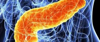
A cyst of the head of the pancreas is a common location in this pathology. Any cystic formation is detected as an ultrasound finding when examining a patient for another reason. In addition to the head, a cavity with fluid is detected in any part of the pancreas. The prevalence of this disease has increased several times in recent years. According to statistics, mostly young people are susceptible to pathology. There is a connection with the increase in the incidence of pancreatitis of alcoholic, post-traumatic etiology. One of the difficulties of diagnosis is that there are no symptoms of pancreatic cysts for a long time. They appear when the cavity reaches enormous sizes. Until this time, the patient can live without knowing that he has a cyst.
What is a pancreatic cyst?
A pancreatic cyst is a fluid formation that develops in the tissue or on the surface of an organ, delimited from the rest of the parenchyma by a capsule. ICD-10 code - k.86.2. It looks like a bubble filled with liquid. To date, there has been no consensus on the definition of pathology. According to some authors, the contents of the cavity are pancreatic secretions, but some believe that necrotic tissue, blood, and inflammatory exudate may be inside the formation. According to certain classifications, the latter relate to complications of pancreatitis and are called pseudocysts.
But in all cases, the conditions under which a cyst forms include:
- tissue damage
- obstructed outflow of pancreatic juice,
- local circulatory disorder.
Causes of pathology
There are several reasons leading to the formation of cavity formations in the pancreas:
- developmental abnormalities (for example, congenital obstruction of the pancreatic ducts),
- acute pancreatitis (85% of cases),
- trauma, when mechanical damage to pancreatic tissue and hemorrhage into the parenchyma occurs (14%),
- helminthiases (echinococcosis, cysticercosis),
- RV infarction,
- blockage of the pancreatic ducts (retention cyst),
- tumors.
Some risk factors lead to the formation of cysts:
- bad habits (alcoholism - 68%, smoking),
- obesity - 33%,
- cholelithiasis (GSD) - 14%,
- diabetes mellitus - 15%,
- surgical interventions,
- unhealthy diet
- calcifications in the pancreas parenchyma.
Classification of pancreatic cysts
According to morphological characteristics, all pancreatic cysts can be classified into:
- true,
- false (pseudocysts) – acquired.
A true cyst is rarely large; therefore, it is discovered infrequently and, as a rule, by accident. Diagnosed in every fifth patient. Its walls consist of an epithelial layer (unlike a pseudocyst, whose walls are fibrous tissue). Due to its small size, there is no compression of surrounding tissues and no external manifestations are observed. If a true cyst is formed as a result of a congenital pathology, a sluggish, unexpressed inflammation and fibrous degeneration of pancreatic tissue is formed: normal pancreatic cells are replaced by connective tissue. In addition, a true cyst can be:
- inflammatory,
- parasitic
- traumatic
- neoplastic.
A pseudocyst is the result of:
- inflammatory diseases of the pancreas (most often pancreatitis),
- injuries.
To designate cavity formations with fluid that arose as a complication of pancreatitis, the Atlanta classification is used and distinguished:
- spicy,
- subacute cyst
- pancreatic abscess.
They differ in the presence of walls and contents:
- in acute cavities, the own walls are not fully formed; their role is played by the tissue of the pancreas itself, ducts, peripancreatic tissue, adjacent organs,
- chronic cysts have their own walls of fibrous cells,
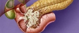
- an abscess is a cavity that appears as a result of pancreatic necrosis or a complication of an existing cyst and is filled with pus.
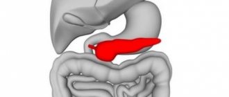
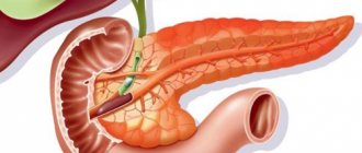
Cystic formations can be located in the area:
- head end
- bodies,
- tail section,
- diffusely occupy the entire organ.
The location of the cyst is possible on the pancreas and inside the organ, in its parenchyma. Also distinguished:
- single,
- multiple cysts.
Reasons for appearance
Most often, false cysts are diagnosed against the background of acute or chronic pancreatitis or alcoholic pancreatitis. In this case, acute or alcoholic inflammation is complicated by a cyst in approximately half of the patients, the chronic form - in 80% of cases.
In the process of cyst formation as a complication of acute pancreatitis, gastroenterologists distinguish 4 stages:
- Stage 1 (1–1.5 months have passed since the onset of pancreatitis) – formation of inflammatory compaction of gland tissue (infiltrate);
- Stage 2 (2–3 months after the onset of pancreatitis) – a cystic capsule begins to form, while its walls are still loose;
- Stage 3 (up to six months) – the cavity capsule is dense, formed by fibrous tissue (mature connective fibers);
- Stage 4 (6–12 months) – the cyst becomes mobile and can be separated from surrounding tissues.
Important! Pancreatic asymptomatic pseudocysts do not always tend to grow; they can shrink and even completely resolve within a year (in 30-40% of cases).
However, cysts larger than 6 cm that have been present for more than 3 months are likely to require surgery - it is only a matter of time.
In addition, the reasons for the appearance of a cystic cavity in the tissues can be:
- hemorrhages in the gland tissue of a non-traumatic nature, accompanied by hematomas;
- injuries of the pancreas and its ducts are accompanied by the release of blood and pancreatic secretions outside the organ. Post-traumatic pseudocysts also begin with a hematoma;
- abdominal or laparoscopic surgical interventions performed during the treatment of cholecystitis or pancreatic tumors. Such pseudocysts are called iatrogenic;
- taking enzyme inhibitors during the treatment of chronic pancreatitis;
- atherosclerosis of the vessels of the gland, provoking circulatory disorders and the appearance of a focus of fibrosis. An uncommon cause of cyst development;
- congenital anatomical anomalies (in children) – we are not talking about congenital cavities, but about the congenital prerequisites for their appearance;
- a tumor process in the pancreas is very rare.
Thus, among the main prerequisites for the development of pseudocysts are pancreatitis and cholecystitis.
Symptoms of cystic formations on the pancreas
The clinical symptoms of an existing cyst in the pancreas depend on:
- parameters,
- location,
- reasons for its formation.
Among the most common symptoms of pancreatic cysts are:
- painful,
- dyspeptic,
- compression of tissues of neighboring organs.
A cyst with a diameter of up to 5 centimeters exists asymptomatically, since it does not disrupt the structure of neighboring organs or nerve trunks. The patient does not even feel discomfort and is not aware of the pathology.
Pain
A large cyst is accompanied by pain. The symptom is characterized by a clear interval: well-being improves for a short time after developing acute pancreatitis or injury.
A pronounced pain symptom appears during the formation of a pseudocyst during pancreatic necrosis and may be associated with a destructive process in the tissue. Gradually the pain decreases, but discomfort remains. In combination with medical history (history of trauma or pancreatitis), this allows one to suspect a cystic formation.
If the pain intensifies, hyperthermia and intoxication appear, you can think about rupturing the cyst.
As the cavity enlarges, the solar plexus is compressed. Intense pain occurs, radiating to the lower back, intensifying even with the pressure of tight clothing. They can only be relieved with narcotic algesics. It decreases when lying on your side with your knees pulled up to your stomach.
Dyspepsia
A pancreatic cyst can manifest itself as dyspepsia, which is expressed by:
- nausea,
- vomiting (at the end of a painful attack),
- unstable stool - constipation, diarrhea,
- flatulence.
If the formation reaches a significant size, the exocrine function of the pancreas decreases due to damage to a large number of cells that produce pancreatic juice with enzymes. This leads to impaired absorption of nutrients in the intestines and weight loss, even if a person eats enough food.
Compartment syndrome
If the cyst is large in size, especially if the cyst is externally located on the surface of the pancreas, nearby organs are compressed:
- when exposed to the portal vein, swelling of the legs, feet,
- when the ureter is compressed, the outflow of urine is disrupted, urinary retention occurs,
- with pressure on the intestinal loops, partial obstruction develops,
- the stomach is compressed by a large cyst in the head, obstructive jaundice develops with its manifestation in the form of icterus of the skin and sclera.
Jaundice, accompanied by painful itching, worse at night, acholic feces, dark urine.
Stages of the disease and symptoms
The ultrasound method allows us to understand what it is, like an X-ray, capable of penetrating any dense organ (including the pancreatic gland) with studying radiation, giving a detailed picture of its internal structure.
It is unthinkable to identify a pseudocyst of small diameter by any other method - it does not make itself felt until its enlargement causes symptoms indicating a serious pathology of the organ containing it.
Considering that almost any damage to the pancreas is caused by alcohol, overeating (eating hastily, dry food, in huge volumes, without thorough chewing and comprehension), stress (including an attitude towards food that is close to a small war with oneself), it is expressed by indigestion in person:
- pain of a periodic nature, involving the epigastric zone (upper third of the abdomen);
- diarrhea;
- belching;
- nausea;
- swelling of the abdomen (to the extent that the diaphragm is pushed up from below, causing shortness of breath and heart attacks).
Obviously, everything the patient experiences is attributed to either poor quality food, an alcoholic episode, or a rush job at work.
Considering that few people think of going to the doctor for such “trifles,” everything is limited to taking No-shpa. Or, having heard a diagnosis of “chronic pancreatitis” from a specialist, the patient continues his previous lifestyle (remembering to take prescribed medications only periodically).
As a result of a chronic burn with alcohol (when it is thrown into the main ductal network of the gland), several reasons arise at once, leading to the bricking up of individual ducts (from which a pseudocyst can gradually form):
- desquamation of the inner layer of cells – the epithelium with the formation of a cell-protein “plug”;
- the formation of a fibrous scar (after repeated inflammation), narrowing the tube that removes juice;
- formation of a tumor that performs the same role.
Considering the close location of the outlet openings of the common bile duct of the liver and the similar pancreas, it is no less common for bile sand and even small pebbles from the bile to be thrown into the ducts of the latter.
Finally, the cause of compression may be hemorrhage into the gland tissue (due to abdominal trauma or for another reason).
Later signs include:
- vomiting due to a disorder in the movement of food through the hollow organs due to a narrowing of the passage from the stomach to the duodenum;
- jaundice due to compression of the common bile duct;
- palpation determination of excess size and mass of the gland;
- weight loss.
When a formation forms inside the small peritoneal (peritoneal) sac, they speak of a peritoneal pseudocyst (with its localization under the layer of peritoneum covering the pancreas in front and above), the consequences of which do not differ from the same formation located inside the organ.
In addition to the presence of the usual and inexpressive symptoms of chronic pancreatitis, manifestations of a pseudocyst that has reached a significant diameter can give a picture of an acute abdomen with:
- its rupture;
- the formation of fistulas connecting the gland with the stomach, pericardium (pericardial sac), and intestines;
- suppuration of the gland tissue - the formation of an abscess, which is dangerous both in itself and fraught with internal gastrointestinal bleeding due to damage to the main vessel - most often the gastric, splenic arteries or duodenal arteries.
In addition, pleural effusion may appear (usually in the left half of the pleural cavity).
Due to the fact that the contents of the described pathological cavity is pancreatic juice with high digestive and destructive properties, its entry anywhere inside the body is a catastrophe no less serious than a heart attack or stroke (when it spills into the pericardial cavity, the development of cardiogenic shock occurs, with infection of the peritoneum - peritonitis).
Video lecture about pancreatic cysts:
Diagnosis of pathology
Laboratory test data for diagnosing a cyst are nonspecific and therefore not very informative. Changes in tests indicate inflammation in the parenchyma. Therefore, the main methods for detecting pancreatic cysts are functional:
- plain radiography of the abdominal cavity,
- Ultrasound – determines the size, number of formations, presence of complications,
- MRI, CT - more accurately assess the parameters and relationship of the cyst with the ducts and surrounding organs,
- scintigraphy (using radionuclides) – clarifies the location of the cavity in the gland tissue,
- ERCP (endoscopic retrograde cholangiopancretography) shows in detail the structure of the formation and the connection with the main ducts, but its implementation is dangerous due to infection.
Plain radiography reveals displacement of any organ from its normal location. This is regarded as an indirect sign of a huge cavity with fluid in the pancreas. You can also find a small stone in the area of former necrosis after pancreatitis.
Laboratory diagnostics
Laboratory studies are not of particular value, since they do not characterize the cystic formation in any way. With their help, the presence of inflammation or a violation of endocrine function (hyperglycemia) is detected as a result of a long-existing cyst in the tail.
General analysis, biochemical parameters of blood and urine, sugar are studied. Pathology reveals increased ESR, leukocytosis, high bilirubin and its fractions, amylase, and increased alkaline phosphatase.
Instrumental diagnostics
Effective methods for detecting pancreatic cysts are ultrasound and CT.
Ultrasound is an accessible and safe method that allows diagnostics to be carried out without special training or complications. Even a child of any age can easily tolerate it, so the study received good feedback from doctors of all specialties.
In many cases, you can get by with the results obtained during ultrasound - this is a screening method that is prescribed first. You can determine the category of the detected formation: is it liquid (cyst) or dense.
The study protocol includes a detailed description of the cyst:
- sizes,
- localization,
- prevalence,
- connection with ducts or other organs and vessels.
It contains conclusions indicating what the results may mean.
For a more detailed examination, if after an ultrasound the diagnosis remains unclear, an MRI or CT scan is prescribed (if there are contraindications to MRI). These methods are much more sensitive, detect cavities from 2 cm, make it possible to see the organ under study in a three-dimensional image, and indicate in detail the connection of the cyst with vessels and other organs.
Plain radiography, in addition to displacement of organs, reveals calcifications or individual foci of calcium salts even in the wall of the cyst.
ERCP is a dangerous method, since contrast injected into the Wirsung duct in a direction that prevents the flow of pancreatic juice causes an exacerbation of the inflammatory process.
Angiography is rarely used, but in complex cases it makes it possible to clarify the nature of changes in the tissues of the gland.
Invasive methods
If the diagnosis is unclear, invasive (violating the integrity of the skin) manipulations are used:
- ERCP is used to examine the gland ducts (choledochus and Wirsung's) and take material for biopsy. There is a high probability of contamination of the body with cancer cells if they are detected.
The method establishes the connection between cavity formation and the pancreatic duct. Consists of sequential endoscopic and x-ray examinations. First, an endoscope with special lateral optics is inserted into the duodenal lumen and fed using a contrast catheter through the orifice of the papilla of Vater. X-ray equipment is then used to take pictures. Based on them, the diagnosis is verified. If necessary, using this method, a biopsy specimen is taken for study. But most often, material is collected percutaneously with a needle or using a biopsy gun.
- Laparoscopy is based on examining the organ with a device with optics, which is inserted into the abdominal cavity through an incision (its approximate diameter is 0.5-1 cm) in the abdominal wall. Simultaneous tissue sampling is possible for further histological examination and differential diagnosis. To do this, an additional incision is made to insert another instrument.
- A biopsy is performed to clarify the structure of the formation, confirm or exclude malignancy.
Diagnostics
If you suspect a pathology of the pancreas, you should contact a physician, gastroenterologist or surgeon. After analyzing the complaints and medical history, the doctor will conduct a general examination. At this stage, it is sometimes possible to suspect large cysts - they are palpated in the upper abdomen as a dense formation.
Additional studies will be required to confirm the diagnosis:
- ultrasound scanning of the abdominal organs;
- computed tomography or magnetic resonance imaging.
According to indications, abdominal radiography and fibrogastroduodenoscopy (FGDS) are additionally prescribed. They are needed to identify concomitant pathologies of the stomach, intestines, liver, and gall bladder. FGDS finds indirect signs of pancreatic disease: erosion of the gastric mucosa at the site of compression by the cyst, esophageal varices, metastases. Biochemical studies (lipase, amylase, etc.) are needed to determine the enzymatic function of the organ and diagnose an acute inflammatory process.
Treatment of pancreatic cyst
Asymptomatic cavity formation is not treated surgically. With a true cyst, there is a possibility that it will resolve. This occurs after the disappearance or reduction of the source of its occurrence (inflammation, degenerative process), even if no therapeutic measures were taken.
For multiple cystic formations, drug treatment is ineffective. You can get rid of them, regardless of the area of localization, through surgery. Surgery is not provided for identified single cysts up to 3–5 cm, if they are asymptomatic. But if signs of malignancy are detected, even a small cyst should be removed to prevent metastasis. Otherwise, in the future it may threaten death at any stage of its development.
The prognosis for a detected cyst is not very encouraging, since it is almost impossible to avoid surgical intervention.
Conservative treatment
Therapeutic treatment of cystic formation is aimed at correcting the underlying disease of the pancreas and is carried out in the presence of:
- single education,
- clear outline
- sizes up to 2 cm.
In all other cases, surgical methods are used.
Conservative treatment consists of prescribing symptomatic therapy for existing inflammation in the gland itself or gallbladder, stomach, duodenum. Aimed at reducing pain, reducing the secretion of pancreatic juice with enzymes. For this purpose the following are used:
- PPIs – proton pump inhibitors (Pantaprazole, Omeprazole),
- enzyme replacement therapy (Creon, Mezim-forte),
- antispasmodics (No-Shpa) and analgesics.
The use of enzymes is necessary for normal digestion and reducing the functional load on the pancreas. If carbohydrate metabolism is impaired, hypoglycemic drugs are used. All medications are prescribed by a doctor, each tablet is selected individually.
Treatment of true cysts
If a true cyst with a diameter of 3-5 cm is verified, which proceeds without complaints or symptoms and does not provoke complications, conservative treatment is applied, and a recommendation is given on the use of diet. The patient is observed for 1.5 months. If the cyst does not grow during this time, the diet is adjusted and the need for an ultrasound scan 1-2 times a year is explained.
A formation with a diameter of more than 6 cm is treated surgically, since over time its size increases and various complications are provoked.
Treatment of false cysts
An identified pseudocyst up to 5 cm in size in the presence of acute pancreatitis does not cause any complaints and can resolve on its own. The approximate duration of this process is 3-4 months.
If a false cyst is observed during a chronic inflammatory process in the gland, it very rarely disappears on its own. Its maximum size is 40 cm. With such parameters, it is possible for it to grow into blood vessels, which leads to fatal bleeding. Therefore, surgical treatment methods are used:
- Removal of a cyst with part of the pancreas.
- Marsupialization (from Latin marsupium - bag): the cyst is dissected and cleaned, and the edges of its wall are sutured into the edges of the abdominal wall incision. Subsequently, the former cavity of the formation is granulated, and a scar is formed in its place. The operation is an invasive treatment method. In recent years, it has been performed infrequently due to prolonged healing and the high likelihood of fistula formation. It is used for echinococcosis or in the case of a large pseudocyst adherent to neighboring organs, when its removal leads to damage to these organs.
- Enucleation (husking).
Surgical methods of treatment
Depending on the severity of the condition and disorders in the pancreas tissue, surgical treatment is performed. Indications for surgical intervention are:
- cysts exceeding 6 cm,
- the presence of malignancy processes,
- penetration of the pseudocyst into the abdominal or pleural cavity,
- suppuration.
For surgical treatment, 3 methods are used:
- resection,
- drainage,
- laparoscopy.
If the abdominal or pleural cavity is affected, the method of external drainage of the cyst and affected cavities is used. The disadvantage of this method is the high risk of developing pancreatic fistula.
If suppuration of a cystic formation is detected, emergency intervention is performed. During the surgical procedure, the cavity is washed and drainage is removed.
Internal drainage of the cavity with fluid is carried out by creating an anastomosis between the cyst and the stomach or intestine. This method is safe and physiological, significantly improves the patient’s well-being - this ensures the removal of the contents of the cyst, relieves pain, and reduces the possibility of relapse.
Cystoenterostomy is a type of surgical treatment: the cyst cavity is opened and emptied with treatment with 96% ethyl alcohol. Then an anastomosis with the intestine is created.
If the cavity is huge, it is resected along with the adjacent part of the organ. The amount of tissue removed depends on the size of the cyst and changes in the tissue.
Minimally invasive surgeries
Minimally invasive interventions are becoming increasingly important in treatment. However, despite the promise of these methods, they often lead to the development of complications.
The laparoscopic method consists of making 3-4 small incisions on the abdomen, after which the operation is performed under the control of the image on the screen from the laparoscope using manipulators that are inserted through the holes. Laparoscopy is used to remove single large cysts. During this procedure, infection of the abdominal cavity and wound is excluded. Due to small incisions, postoperative hernias do not form. The patient experiences virtually no pain, so strong narcotic drugs are not prescribed; ordinary analgesics are sufficient. The recovery period takes a short period of time: discharge from the hospital occurs in a maximum of a week.
In recent years, new technology for the treatment of NOTES has been developed. It allows you to remove cystic formations in the pancreas through access through the natural openings of the body. When performing it, not a single incision is made. Due to the high cost of equipment, this treatment is carried out in only a few clinics. In our country, such equipment is available in a clinic in Novosibirsk.
Abdominal operations
Sometimes it is necessary to treat cavity formations in an open way - using laparotomy. These are difficult abdominal operations involving a large incision on the abdominal wall. This access is performed when a large volume of surgery is required to remove part of the pancreas or the entire organ:
- distal resection of the pancreas (in the presence of large cavity formations in the body and tail),
- drainage of suppuration,
- complete pancreatectomy,
- removal of the head.
Indications for complete resection of the pancreas are multiple cavities with fluid throughout its entire length.
Surgical intervention
If the pseudocyst grows to a large size (more than 6 centimeters), does not resolve on its own, and conservative treatment does not bring results, then a decision is made on surgical intervention.
Surgical removal can be different:
- Percutaneous drainage. It is considered one of the most effective methods. During the operation, drainage is installed through the skin and wall of the gland. Doctors sometimes use this method with caution, as some patients may experience certain complications.
- Linear endoscopic echography. With this method, the pseudocyst is drained through the stomach or intestines of the person. The method is also considered effective, but it can only be carried out if the formation is located in close proximity to the stomach.
- Transpillar drainage of pancreatic pseudocyst. This method cannot be considered a full-fledged surgical one. Its essence is to install a special stent. It is placed into the human body during the next ERCP.
- Internal drainage. It is considered an outdated method. It is practically not practiced in modern medicine due to the fact that many patients tolerate such an operation very poorly.
- Complete surgical removal of the pseudocyst. During the operation, a large incision is made in the abdominal cavity. This method is very traumatic, but it is very often used when the formation is located in the head or tail of the pancreas.
Before any surgery, the patient must follow a strict diet.
Traditional methods of treatment
Many patients independently use various methods of traditional medicine to treat cystic cavities in the pancreas, believing that they can stop further growth of the formation and prevent the appearance of new ones. For this purpose, various medicinal herbs (celandine, immortelle, chamomile), propolis, and mumiyo are used. It must be remembered that the main condition for treatment with any folk remedies, even the seemingly most harmless ones, is a preliminary consultation with a doctor. The pancreas is a delicate and extremely sensitive organ, so the doctor must evaluate the balance of benefits and harmful effects. For example, propolis - “bee glue” - cannot be used independently without the consent of a doctor, as it can cause a severe allergic reaction. When taking it, it is necessary to adhere to a strict dosage and duration of use, even for people who are not prone to allergies. Alcohol tinctures, which are prepared from medicinal plants, are strictly contraindicated for any pathology of the pancreas.
Professor I.P. Neumyvakin proposed treating pancreatic cysts at home using soda and hydrogen peroxide, explaining the therapeutic effect by the influence of oxygen released from the peroxide on the body. He considered these remedies universal and compared them in terms of their potency with immunostimulants. Considering the side effects, especially for existing stomach diseases, this method should not be used independently. The claim that it has cured cysts and even cancer is greatly exaggerated.
In no case can such therapy exclude traditional therapy, especially if the cyst needs to be operated on. Considering the high risk of complications of large cystic formations, treatment that is not confirmed by evidence-based medicine cannot be used.
Clinic
Most PCs are asymptomatic, but they can have different clinical manifestations depending on their size and location.
- An enlarged pseudocyst can cause abdominal pain, obstruction of the duodenum, blood vessels or bile ducts. Fistulas with adjacent organs, the pleural cavity, or the pericardium may form.
- Spontaneous infection with abscess formation.
- Digestion of adjacent vessels can cause the formation of a pseudoaneurysm, which can cause a sharp increase in the size of the pancreas or bleeding from the gastrointestinal tract as a result of bleeding into the pancreatic duct [3].
- Pancreatic ascites and pleurisy can form when the pancreatic duct ruptures with the formation of a fistula with the abdominal or thoracic cavity or when the PC ruptures.
Diet for pathology
If there is a cystic formation in the pancreas, proper nutrition is part of the treatment. Dietary table No. 5 according to Pevzner is prescribed. It excludes the consumption of fatty, spicy, fried, smoked foods. Food should be warm, crushed, and you should eat small portions 4-6 times a day. This will reduce the functional load on the pancreas and avoid tissue damage with the formation of a cyst or its further growth. The duration of the diet, the list of prohibited foods, and a specific menu will be prescribed by the doctor, taking into account the clinical situation. Dietary restrictions are observed for several months, sometimes they have to be observed throughout life.
Stages of formation
Based on their location, false cysts are divided into formations of the head, tail or body. In addition, they can be postoperative, post-traumatic and pancreatic.
There are several stages of formation of neoplasms:
- Initial. It takes about one and a half months and is characterized by the formation of a cyst cavity.
- Second. The stage can last about three months. In this case, new loose tissue develops in the formed area.
- Third. Characterized by the completion of the formation of a fibrous capsule.
- The last one. The capsule becomes denser. The process of its formation is completed.
Pancreatic pseudocysts: what are they and how can they be located? False cysts can be single or present in large numbers in the organ area. A pathology is considered acute if it remains in the organ for three months, subacute - about six months and chronic - more than this period.
Prognosis after treatment and prevention of pancreatic cyst
The prognosis for an existing pancreatic cyst depends on:
- reasons for development,
- timeliness of diagnostic measures,
- method of surgical treatment used.
Prevention comes down to avoiding the consumption of alcohol-containing drinks, timely adequate treatment of pathologies of the digestive organs, and a balanced diet.
Consequences and complications of cysts on the pancreas
If not detected in a timely manner, a cavity formation of a solid size poses a danger due to complications, which, according to statistics, account for 10-55% of all cases in the form of:
- suppuration,
- perforations,
- bleeding,
- fistulas,
- peritonitis.
If it is not treated in a timely manner, severe consequences may develop:
- the cyst may burst:
- education becomes malignant.
Even if the cyst is removed surgically, there remains a risk of recurrence.
Possible complications
If treatment is not started in a timely manner, a complication of the pathology cannot be ruled out. Some of these complications include:
- Cyst rupture. Occurs rarely, which is facilitated by trauma to the pancreas.
- Suppuration of the cyst.
- Bleeding in the cyst cavity.
It is possible that complications may occur during surgical interventions. These complications include:
- The occurrence of extensive hemorrhages.
- Violation of the integrity of the mucous membrane of other gastrointestinal organs.
- The appearance of scars.
- Perforation of the cyst.
- Infection.
Treatment in clinics in Russia and abroad
Great successes in the treatment of pancreatic cysts have been achieved in Israel, Germany, and the USA. The high level of development of medicine and modern medical technologies make it possible to identify this pathology at the earliest stages and successfully treat it without complications. Only minimally invasive methods are used for treatment. In Israel, a DaVinci robotic installation is used, which performs the most delicate manipulations with a hard-to-reach cyst localization in the pancreas.
In Moscow, treatment of pancreatic cysts is professionally carried out in specialized institutions: Research Institute of Emergency Medicine named after. N.V. Sklifosovsky, Institute of Surgery named after. A.V. Vishnevsky. To avoid tragic consequences, it is important to consult a doctor when the first signs of the disease appear.
Bibliography
- Vinogradov V.V. Tumors and cysts of the pancreas. M.: Medgiz, 1959.
- Bozhenkov, Yu. G. Practical pancreatology. Guide for doctors M. Med. book N. Novgorod Publishing House NGMA, 2003
- I.N. Grishin, V. N. Grits. Cysts, fistulas of the pancreas and their complications. St. Petersburg, Higher School, 2009
- V.A. Kubyshkin, G.G., Karmazanovsky, S.A. Grishankov. Cystic tumors of the pancreas: diagnosis and treatment. M., Vidar-M, 2013
Diagnostic detection of pathology
Diagnosis of the disease is based on the collection of medical history data, as well as the presence of pancreatic diseases in the patient’s history. These can be types of ailments such as pancreatitis, tumors and diabetes. If the patient smokes or abuses alcohol, then the likelihood of developing pancreatic pseudocysts is quite high.
When examining a patient, the doctor pays special attention to the clinical picture of the disease. If the patient experiences abdominal pain, as well as nausea and vomiting, the doctor may suspect the development of a tumor in the pancreas. In addition, attention is paid to laboratory research. These studies include the following procedures:
- Amylase. If pathology is present, the normal value of this indicator will differ by 50%.
- General blood analysis. If an increase in leukocytes in the blood is detected, then we can talk about infection of the cyst cavity. With a reduced level of hemoglobin and red blood cells, it can be judged that bleeding is observed from the pseudocyst.
- Electrolytes, creatine and glucose. If the patient has signs of a pseudocyst, then hypocalcemia, hypokalemia, and hypomagnesemia will be detected.
We also recommend viewing: Causes, symptoms and non-surgical treatment of stones in the pancreas
However, it is important to emphasize the fact that laboratory testing does not make it possible to make a 100% diagnosis of pancreatic pseudocyst. If a specialist has doubts about the correctness of the diagnosis, then one of the following instrumental diagnostic methods is prescribed:
- CT scan. One of the most popular and sought-after diagnostic methods, which is indicated for the slightest suspicion of a tumor. The advantage of this method is the relatively low cost of diagnosis.
- Ultrasound examination or ultrasound. Another relevant diagnostic method, with which you can carry out primary diagnosis, as well as carry out dynamic monitoring of the development of a pseudocyst that was detected early. The ultrasound technique is also an inexpensive research method, therefore, in the process of treating pathology, it can be prescribed for use in any quantity.
- Angiography. The technique is carried out mainly when there is a suspicion of bleeding from a pseudocyst, as well as for the differential diagnosis of other ailments.
Based on the above diagnostic methods, a diagnosis is made, on the basis of which treatment is carried out. Let us consider in more detail what the treatment of pathology includes.