General information and statistics
A calculus is a dense stone that forms in the cavitary organs and excretory ducts of the glands. Kidney stones are formed due to impaired filtration in the kidneys and metabolism in the body.
The pathology occurs among both children and adults. Mostly men are susceptible to the development of this pathology, although this disease also occurs in women.
According to statistics, in the world 7% of men and 3% of women suffer from urolithiasis. In Russia, every second patient hospitalized in the urology department has this diagnosis.
Separation of stones by size
When identifying kidney stones, it is very important to assess their number and size. Multiple fine sand (the size of each grain of sand is up to 1 mm) is the best option for treatment. It is enough to change your diet, start drinking special mineral water, and the risk of stone formation will disappear.
It is more difficult to choose treatment when detecting single or multiple microliths, the size of which does not reach 10 mm. Considering that the ureter leaving the kidney has a diameter of 6–8 mm, spontaneous passage of these stones can be expected. However, the risk of an attack of renal colic due to the fact that several small kidney stones get stuck somewhere in the ureter is quite high. Therefore, one must be extremely careful in approaching any methods of removing kidney stones if the estimated size of at least a few of them exceeds 8 mm.
If stones larger than 10 mm are detected, which cannot move into the ureter and spontaneously pass, an individual approach to treatment is required. Large kidney stones larger than 100 mm are removed surgically.
Pathogenesis and causes of the disease
To understand the mechanism of kidney stone formation, you need to analyze the composition of urine. Human urine is a liquid containing a rich variety of chemicals.
Urine is saturated with crystals, but if a person’s kidneys are functioning fully, they do not precipitate, and this is all thanks to the protein compounds contained in the urine. Proteins prevent crystals from sticking together and settling.
If mucoproteins, for example, pus or mucus, appear in the urine, they neutralize the effect of protein compounds that prevent crystals from sticking together, which is why the formation of a future solid formation begins.
It gradually increases due to the addition of salts, fibrin threads and foreign bodies and interferes with the normal functioning of the kidneys.
The essence of the problem is that stones move into the urinary tract, thereby preventing the full functioning of the urinary system.
The causes of urolithiasis include:
- untreated foci of inflammation in the organs of the excretory system;
- diseases that disrupt mineral metabolism, such as gastritis, liver failure, osteoporosis;
- hereditary factor;
- violation of the drinking regime;
- excess weight;
- poor nutrition;
- congenital kidney anomalies.
It is believed that excessive consumption of foods containing animal protein and oxalic acid increases the risk of urolithiasis.
CONCRETE
CONCRETE
(lat. concrementum pile, accumulation; synonym:
stones, accretions
) - dense, rocky formations found in the human and animal body. Most often, this term defines formations that arise in the cavities of organs or their ducts: in the bile and urinary tracts (see Gallstones, Urinary stones), in the ducts of the salivary glands (see Sialolithiasis), pancreas, in the intestines, in the bronchi (see Broncholithiasis), in the nasal passages (see Rhinoliths), in the crypts of the tonsils, in the veins (see Vein stones), in the cavities of the peritoneum and pleura, the vaginal membranes of the testicles, in the joints, etc. This term is also used in relation to calcareous deposits in tissue, for example, to petrification in the area of a tuberculosis focus or infarction, at the neck of teeth (see Tartar). Fecal stones (see), fibrin convolutions in the renal pelvis are also sometimes called K.
K.'s education is mainly due to physical and chemical. and fiziol, properties of the environment. Phys.-Chem. factors for the occurrence of K. are a violation of the solubility of organic and inorganic compounds that are suspended or dissolved in liquids (urine, bile, glandular secretions) or in tissues, which is facilitated by the so-called. protective colloids, for example, bile acids that retain cholesterol esters in the solution. When changing physical-chemical properties of the solution, which leads to an imbalance in the system; substances dissolved or suspended in the liquid precipitate. Fiziol, the factors causing K.’s formation can be local and general. General factors include metabolic disorders, which can be congenital or acquired, associated with a person’s diet and lifestyle, with destructive processes, with intoxications, etc. Thus, the processes of formation of blood cells in the urinary and biliary tracts are facilitated by a sedentary lifestyle , nutritional characteristics, feverish conditions, climatic factors. Among local factors, disturbances in the secretory and resorptive activity of the organ, stagnation of secretions, and inflammatory processes play an important role in the process of formation of blood cells. When the processes of secretion and reabsorption are disrupted, the concentration of dissolved substances increases and they fall out of the solution. Inflammatory processes contribute to stagnation of the contents of hollow organs and ducts, which leads to precipitation, changes in salt solubility and disruption of the protective colloids.
In cases where the processes of stone formation are caused by metabolic disorders, we are talking about metabolic K., and when inflammatory changes prevail, we talk about inflammatory K. In the urinary tract, sometimes there are medicinal K., formed when using sulfonamide drugs, which, excreted by the kidneys, crystallize, which is especially favored by the acidic or neutral reaction of urine.
K. have different sizes, shapes, structures and chemicals. composition, depending on the place of origin and mechanisms of formation. Some cells can be visible only with the help of a microscope (microlites), others reach significant sizes (macrolites). K.'s shape often follows the contours of the container where they are formed. Thus, stones in the gall bladder and bladder are round or ovoid in shape, in the excretory ducts they are cylindrical in shape. There are single (solitary) K. and multiple. The surface of K. can be smooth or rough. In the case when multiple concretions are closely adjacent to each other, numerous edges and areas—faceted concretions—are formed on their surfaces along the line of contact. Concretions can be very hard or less hard—the consistency of chalk or compacted sand.
In K., a core and layers are distinguished. The core (matrix) of each K. is an organic or colloidal substance, on which inorganic substances are layered. The colloidal base of the cell consists of mucoproteins and polysaccharides and can be presented with desquamated epithelium, leukocytes, thickened mucus, protein sediments, fibrin, bacterial accumulations, etc. Layering characterizes periods of cell growth due to the salts precipitated in this colloidal system. In some cases, some metals and enzymes contained in the colloidal system can act as catalysts in the formation of such deposits (for example, copper in the formation of gallstones). Sometimes the kernels of cells consist of one substance, and successive layers have a different composition—mixed cells, for example, cholesterol-pigment-calcareous cells in the gallbladder. The difference in the composition of this kind of combined salts is associated with changing conditions of salt precipitation, which, in turn, depends on complex and varied changes in a given cavity. The layered structure of crystals is characteristic of colloids, while radially arranged stripes are characteristic of crystalloids.
The composition of K. depends on the substances present at the sites of their formation. Thus, K. of the biliary tract consist of bile pigments, cholesterol and calcium salts, and K. of the urinary tract can contain uric acid, uric acid salts, calcium oxalate, calcium phosphate, less often calcium carbonate, cystine, xanthine. Sometimes K. are represented by one substance, for example, cholesterol gallstones, in other cases K. consist of protein substances that form the colloidal base on which salts precipitate—colloid-crystalloid K.
In some cases, K. does not cause a wedge, symptoms and are discovered by chance. K. are often accompanied by painful manifestations. Getting into narrow channels (bile ducts, ureter), they cause blockage, accompanied by pain, inflammation, and sometimes necrosis and perforation of the wall with the formation of fistulas. Under the pressure of fluid accumulating in the cavities, the surrounding tissues undergo atrophic and sclerotic changes. In the kidneys this leads to hydronephrosis, less often to fatty degeneration, in the liver - biliary cirrhosis, in the pancreas - to atrophy of the excretory parenchyma. The complex of clinical and morphological changes accompanying the presence of K. makes it possible to identify independent diseases characterized by a certain wedge, picture (see Gallstone disease, Kidney stone disease) and requiring conservative or surgical treatment.
Bibliography:
Avtsyn A, P. Introduction to geographical pathology, p. 226, M., 1972; Borisova-Khromenko V. M. Urolithiasis in the Altai Territory and some of its features, Barnaul, 1973, bibliogr.; Weinberg Z. S. Kidney stones, M., 1971, bibliogr.; Galeev M. A. Gallstone disease, Ufa, 1975, bibliogr.; Davydovsky I.V. General human pathology, p. 148, M., 1969; Kiseleva A.F. and Blagodarov V.N. Pathological anatomy and pathogenesis of kidney stones, Arkh. pathol., t. 36, no. 8, p. 3, 1974, bibliogr.; Novikov I. F. Ureteral stones, L., 1974, bibliogr.; Lehrbuch der speziellen Pathologie, hrsg. v. LH Kettler, S. 533, Jena, 1976; Walter JB a. I srae 1 MS General pathology, Edinburgh, 1974.
G. M. Mogilevsky.
Types of stones
Depending on the chemical composition, there are several types of kidney stones, differing from each other in the reasons for their formation, chemical composition and size, namely:
- Urate. These are smooth, rounded formations. The size varies from one millimeter to several centimeters. Formed from uric acid and salts. The urates themselves are yellowish-brown in color. If untreated, urates turn into coral-shaped formations that can spread to the entire renal cavity.
- Oxalate . They are the most common among other types of kidney stones. Oxalates are formed due to excessive amounts of oxalic acid and a disrupted process of its metabolism in the body. They are deposited mainly on the kidney calyxes and take the form of plaques of different sizes (from 1 millimeter to 4 centimeters). Oxalates are considered the most dangerous kidney stones because they damage the kidney tissue, leading to bleeding.
- Struvite . These solid neoplasms are also called infectious because they develop against the background of inflammatory processes in the kidneys. Consist of ammonium phosphate and calcium carbonate. They are formed due to stagnation of urine and infection. The danger of struvite formations is that they can provoke acute renal failure and sepsis.
- Phosphate. They consist of salts of phosphate acid and rapidly increase in size, which can lead to filling of the hollow structures of the kidney. In this condition, a person immediately undergoes surgery to remove the stones.
- Protein . These stones contain fibrin and bacteria. Their size does not exceed a few millimeters. They are considered the least common among other types of stones.
- Cholesterol . They arise due to a violation of cholesterol metabolism in the body. The formations are soft, but black in color. Easily destroyed.
- Cystine . They are formed against the background of a rare kidney pathology - cystinuria, which manifests itself in the fact that cystine is not reabsorbed, but accumulates in the organ.
- Xanthinaceae . They develop against the background of a congenital abnormality of kidney function. Stones form because xanthine is excreted from the kidney but is not converted into uric acid.
The danger of kidney stones is that their rapid increase leads to blockage of the urinary ducts and complicates the removal of urine from the body.
General rules and methods of treatment
The main goal of treatment is to remove stones and eliminate the symptoms of nephrolithiasis. Depending on the size of the formations, their location in the kidneys, and chemical composition, conservative and surgical treatment may be offered.
Medications
When renal colic develops, painkillers and antispasmodics are prescribed:
- Diclofenac;
- Nimesil;
- Codeine;
- Papaverine;
- alpha1-blockers;
- opioid analgesics for unbearable pain.
Often, with renal colic, it is necessary to place a novocaine blockade to relieve pain.
If there is a urinary tract infection, antibiotics and uroseptics are prescribed. The most effective antibiotics are from the group of cephalosporins, macrolides, and fluoroquinolones.
They help remove small stones, as well as provide an antiseptic effect with plant-based uroseptics. They are also used to prevent the growth of stones:
Some types of small formations in the kidneys are tried to be dissolved using medications:
- Potassium citrate;
- Tiopronin;
- Litostat and others.
Dense stones, such as oxalates, do not dissolve and drug therapy is ineffective against them.
Find out about the normal level of bilirubin in urine, the reasons for the deviation and options for correcting the indicators.
What to do if your kidneys hurt badly during pregnancy? Read the answer in this article.
Go to https://vseopochkah.com/bolezni/drugie/glomerulonefrit-u-detej.html and read information about the causes of acute kidney glomerulonephritis in children and methods of treating the disease.
Surgery
Surgery remains the most effective treatment for nephrolithiasis today. The choice of surgical intervention method will depend on the size of the stone, its location and access.
The following types of surgical treatment of deposits are used:
- Extracorporeal lithotripsy is the crushing of formations using a special device using ultrasound, laser and other types of waves. The operation does not require cutting the skin; crushing of kidney stones is carried out remotely.
- Contact lithotripsy is performed using an endoscope. The instrument is inserted from the lower part of the ureter into the bladder, then to the immediate location of the stone. Crushing is carried out by exposure to certain shock waves.
- Open surgical removal is carried out in cases where the stones have reached an impressive size and the location of the deposits does not make it possible to remove them in any other way.
Classification of stones
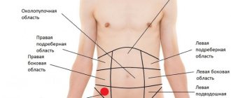
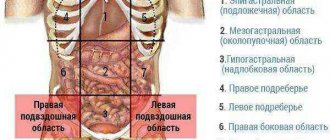
Kidney stones can be classified not only depending on their chemical composition, but also in terms of their location, namely:
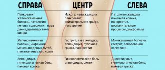
- Right kidney . It is in the right kidney that stones most often form. Blockage of the urinary duct of the right kidney is manifested by nausea, arrhythmia, stabbing pain in the right side, weakness and dry mouth.
- Left . Stones in the left kidney are diagnosed less frequently, and with left-sided lesions, weakness and severe pain in the pit of the stomach are observed.
Diagnosis of stones
Several procedures are necessary for diagnosis. Because the symptoms of urolithiasis and other kidney diseases are similar, careful and accurate diagnosis is required. Another problem is the similarity of the manifestations of left-sided stones with a heart attack, and right-sided ones with appendicitis or problems with the gallbladder and liver. Only an accurate diagnosis will allow you to prescribe the correct treatment.
Diagnostic procedures
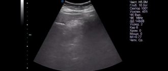
- Kidney ultrasound is the most common, accessible and quite informative technique.
- Computed tomography is a study that allows you to most accurately determine the size and location of stones and identify even the smallest ones.
- Laboratory tests are necessary to detail the clinical picture and assess the patient’s condition.
The primary detection of formations in the kidneys occurs during ultrasound. This is noted in the diagnostic results, which are necessary for the doctor to make a diagnosis and prescribe treatment. Left-sided urolithiasis is not as severe and painful as right-sided one.
If a right kidney calculus is detected, this means that there is a microlith or stone in the right kidney, and it is necessary to find out its nature in order to take timely measures.
If necessary, diagnosis can be supplemented with contrast radiography, MRI, and radioisotope scintigraphy. A study of urine and blood shows the nature of the stones, the condition of the kidneys and the entire body. To identify the infectious process, a urine microflora analysis is done.
Manifestation of the clinical picture
Urolithiasis in 97% of cases is asymptomatic, but until it is complicated by pyelonephritis, cystitis or blockage of the urinary ducts. In 3% of cases, patients initially feel periodic nagging pain in the lower back.
Stages of development
When the patient begins to develop kidney stones, no symptoms are observed. Later, microtraumas to organ tissue provoke pyelonephritis, the symptoms of which include:
- increased body temperature;
- change in the color and smell of urine;
- nausea;
- prostration;
- heaviness and pain in the lower back.
When kidney stones larger than 5 millimeters are evacuated, a person develops renal colic.
With obstruction, sharp pain occurs in the lower back, and it does not depend on the position of the body, which is why the patient becomes restless. Nausea, vomiting, severe intestinal cramps and increased urination are also noted.
If the stone continues to remain in the ureter, the functions of the excretory organs are inhibited. In addition, severe trauma to the ureteral mucosa occurs with stones.
Gradually, the affected organ swells, and the patient suffers from severe pain. If the solid formation is not removed, urosepsis and acute renal failure develop, which can be fatal.
Causes of kidney stones
The process of formation of kidney stones can be influenced by various factors, both congenital and acquired. Congenital causes include genetic predisposition or geographic factors (hot climate).
Acquired reasons:
- chronic and infectious diseases of the gastrointestinal tract, genitourinary tract;
- disturbance of electrolyte metabolism, which is associated with diseases of the parathyroid gland, gout, osteomyelitis, bone tissue destruction;
- lack of fluid intake;
- unhealthy diet (abuse of salty, fried, spicy foods);
- deficiency of vitamins A, D;
- impaired urinary outflow due to narrowing of the urinary tract, adhesions, tumors, prolapse of the kidney;
- long-term use of certain medications (Aspirin, Tetracycline, Biseptol).
Diagnostic measures
If a person experiences symptoms of urolithiasis, he should consult a nephrologist. An accurate diagnosis is made only after the patient has completed all the necessary studies:
- general blood test;
- detailed urine analysis;
- ultrasound examination;
- excretory urography;
- computed tomography.
During the diagnostic process, attention is paid to the presence of diseases that provoke the formation of kidney stones (diabetes mellitus, obesity, coronary heart disease).
Mechanism of stone formation
Human urine consists of water and salts dissolved in it, the main cations of which are chlorides, sulfates, and phosphates. The kidneys take an important part in metabolism, so urine contains protein breakdown products: uric acid and its compounds. The elements that form the solid part of urine enter into various chemical reactions with each other, the product of which is tiny crystals.
Such a saturated solution serves as a favorable environment for solid particles to precipitate, but with an active filtration process, all of them are removed from the body through urination. If the dynamic balance is disturbed under the influence of a negative factor, the following processes occur:
- The content of protein threads in the urine, which prevent the enlargement of crystals, is reduced.
- The content of pathological mucus or pus increases, which, despite their protein origin, provoke the clumping of particles.
- The resulting crystal becomes the basis of the future stone.
Such a nucleus can be formed from foreign bodies, bacteria, cheesy urinary sediment, the protein thread itself, and chemical compounds. The process begins to develop at the apices of the left or right renal pyramids, and the collecting ducts serve as a site for the accumulation of crystalline formations. The surface tension of the microlite is increased; it attracts molecules of chemical compounds and protein components of urine. Over time, small and large stones are deposited on the walls of the papillae, which gradually move down towards the bladder.
Kidney stones can come in a variety of sizes
Therapy methods
The choice of treatment tactics for kidney stones is influenced by the patient’s general health, his age, size and types.
Traditional methods
Conservative therapy for urolithiasis includes taking medications that help relieve inflammation, pain and dissolve solid formations.
Traditional treatment methods are used when the diameter of the formations does not exceed 5 millimeters.
The following drugs help remove stones from the kidneys:
- Cyston;
- Canephron;
- Urolesan;
- Phytolysin;
- Stonebreaker;
- Gortex.
Drugs such as Papaverine, No-Shpa and Baralgin help relax the muscular layer of the urinary ducts and relieve pain associated with the passage of stones.
To dissolve urates, the patient is prescribed Asparkam and Blemaren - drugs that help remove sand. For small oxalates, the patient is prescribed a complex of vitamins A, E and group B.
If a bacterial infection occurs, antibacterial drugs are prescribed - Ceftriaxone, Biseptol.
A therapeutic diet is used to dissolve and remove phosphate stones. If the stones provoke an inflammatory process, the patient is prescribed antibiotics.
Treatment depending on the course
As noted, in the initial stages of urolithiasis, conservative therapy is used.
If there is a risk of obstruction of the urinary ducts due to the rapid increase in stones, modern methods of crushing stones are used. Solid formations are crushed using laser or ultrasound.
Ultrasound lithotripsy includes the following types:
- contact: implies instrumental destruction of stones;
- remote shock wave: using ultrasound, a shock wave is generated that remotely destroys solid formations without damaging the organ;
- extracorporeal: used when the diameter of the stones does not exceed 25 mm, and involves the impact of a burst of shock waves on the stones.
Surgical intervention is used if the size of the stone exceeds 5 centimeters, and previously used treatment methods have proven ineffective.
Surgical removal of a foreign body is prescribed for acute obstruction of the urinary tract, which has caused a sharp disturbance in the outflow of urine.
Kidney transplantation is resorted to in the case of the formation of cystine and xanthine stones, which are formed due to congenital anomalies of the paired organ.
ethnoscience
Non-traditional methods of treating urolithiasis are used after consultation with a doctor. Effective folk remedies for stones include:
- Rosehip root decoction : take 2 tablespoons of phyto-raw materials, pour a glass of water over them and boil for 25 minutes. After this, the broth is infused, filtered and taken three times a day, a third of a glass, for two weeks.
- Collection for removing stones : take 2 tablespoons of a mixture consisting of knotweed, lemon balm, sage and St. John's wort. Pour half a glass of boiling water over everything and insist. During the first week, take half a glass of the infusion, and from the second week, add 5 drops of fir oil to the decoction. The course of treatment is 3-4 weeks.
- Bearberry decoction : pour a tablespoon of dry herb with a glass of boiling water, place the preparation in a water bath, cover with a lid and simmer for 30 minutes. The decoction is taken one tablespoon 3-5 times a day.
Alternative treatment methods cannot be considered as a complete replacement for taking pharmacological drugs for urolithiasis.
Complications
If urolithiasis is not treated, it provokes the following complications:
- Hydronephrosis : complete blockage of the outflow of urine and its accumulation in the kidney, which leads to partial or complete necrosis of the organ.
- Acute kidney failure : A rare condition that occurs when there is bilateral blockage of the urinary ducts.
- Hypertrophic cystitis : develops with constant irritation of the bladder by stone particles.
Complications may be of infectious origin. Examples of such complications include pyelonephritis, chronic cystitis and urethritis.
Treatment options
Optimal treatment methods are selected depending on the size of the stones. If the grains of sand do not reach one millimeter, no specific treatment is required - it is enough to simply adjust the drinking regime to ensure that the deposits are washed out. For small stones (up to five millimeters), specialists can ensure their removal from the kidneys naturally.
Stones larger than five millimeters cannot enter the ureter on their own and require drug therapy. Large crystals (from ten millimeters) pose a serious threat to health. They can be eliminated through surgery.
Surgery
There are several methods for getting rid of large kidney stones; doctors determine which one is suitable for a particular patient. Can be used:
- laparoscopy;
- remote crushing;
- abdominal surgery;
- nephroscopy;
- contact crushing.
During laparoscopy, kidney stones are removed through three small incisions, each no more than five millimeters in size. The likelihood of tissue scarring is minimal, and recovery will only take a few days.
Remote crushing is considered the most gentle option, but the method is used only when necessary to remove stones that are less than two centimeters in size. Dense neoplasms are broken up by laser or ultrasonic waves. They turn sediment into sand, which is then excreted from the body through urine.
Abdominal intervention is performed only in complex cases, if other procedures seem to doctors to be insufficiently effective. The disadvantages include a long and difficult rehabilitation period. With contact crushing, the impact on the stone occurs using special equipment that is inserted through the urinary canal. The stones become smaller and leave the body on their own.
During nephroscopy, a small puncture is made in the lumbar region on the right or left. Through it, a metal tube is inserted into the kidney and removes or destroys the stone. The method is used for single formations and the absence of diseases of the urinary system.
Drug therapy
Conservative treatment is possible for small stones, and only if they do not interfere with the flow of urine. Conventionally, medications can be divided into two types. Some of them are effective against all types of stones, while others are only suitable for getting rid of specific types.
Allopurinol, potassium citrate and Purinol contribute to the destruction of urates. They reduce the acidity of urine, after which the stones gradually dissolve. You must first make sure that the patient is not diagnosed with excess calcium in the urine.
To remove phosphates, the acidity in the urine must be increased. This can be achieved through diet (the diet includes acidic foods) and madder extract. Thanks to the antispasmodic effect of the drug, the process of removing stones through the urinary tract is facilitated.
A universal medicine is Blemaren. Due to its use, the composition of urine changes - it becomes alkalized, the content of potassium and sodium ions increases. The product helps remove existing stones from the kidneys and prevents the formation of new ones. This occurs due to the normalization of acidity levels.
Disease Prevention
To avoid the formation of stones, it is necessary to maintain a drinking regime, monitor the quality of fluid consumed, eat right, control body weight and promptly treat diseases of the excretory system.
Kidney stones are a disease that not only spoils a person’s quality of life, but also poses a threat of developing severe complications. This disease requires timely correct treatment prescribed by a specialist.
If you start therapy on time, the chances of a full recovery increase. Self-medication for urolithiasis can cause irreparable harm to the patient’s health.
Prevention measures
If a person has a tendency to deposit salts and form stones, preventive measures must be taken to minimize the possibility of this process.
Recommendations:
- increase the amount of fluid consumed so as not to create a high concentration of salts in the urine,
- follow dietary rules, limit the consumption of foods that promote stone formation,
- correct metabolic and hormonal disorders in the body,
- to refuse from bad habits,
- promptly treat chronic diseases and inflammatory processes of the urinary organs,
- regularly take tests, conduct ultrasound of the kidneys to monitor the health of the paired organ.
Kidney stones are a serious pathology. If the disease is not treated, serious complications may follow. The sooner deposits are detected, the higher the chance of getting rid of them painlessly. Even complete removal of kidney stones does not guarantee that they will not appear again. It is better to eliminate all prerequisites for this in advance and focus on prevention. At the slightest suspicion of kidney failure, you should consult a nephrologist or urologist and undergo a full examination.
Nephrolithiasis is the most common urological disease, manifested by the deposition of stones in the kidneys. Video - fragment of the TV show “About the Most Important Thing” about the causes, treatment features and consequences of deposits in a paired organ:
Treatment methods
The main types of treatment for kidney stones are:
- conservative therapy aimed at removing sand and small stones up to 6 mm in size;
- a non-surgical method of remote crushing of medium-sized stones with subsequent gradual removal of fragments through the ureters;
- surgical treatment for large stones.
For bilateral disease, the choice of treatment option depends on the size of the stone and the risk of complications. With macroliths on the left and multiple microliths on the right, surgical intervention is performed in stages: first, the stone in the left kidney must be removed, and after 2–3 months, stones in the right kidney can be addressed. A one-time operation is performed extremely rarely due to the high risk of renal failure.
The formation of a kidney stone is a complex and step-by-step chemical process that occurs against the background of accompanying factors and the obligatory accumulation of salt residues in the urine. The treatment option for nephrolithiasis is always selected individually. The chances of avoiding surgery largely depend on the size of the stones.