Chinese sages said that if a person has a healthy intestine, he can overcome any disease. Delving into the work of this organ, you never cease to be amazed at how complex it is, how many degrees of protection are built into it. And how easy it is, knowing the basic principles of its work, to help the intestines maintain our health. I hope that this article, written on the basis of the latest medical research by Russian and foreign scientists, will help you understand how the small intestine works and what functions it performs.
Structure of the small intestine
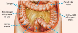
The intestine is the longest organ of the digestive system and consists of two sections. The small intestine, or small intestine, forms a large number of loops and continues into the large intestine. The human small intestine is approximately 2.6 meters long and is a long, tapering tube. Its diameter decreases from 3-4 cm at the beginning to 2-2.5 cm at the end.
At the junction of the small and large intestines there is an ileocecal valve with a muscular sphincter. It closes the exit from the small intestine and prevents the contents of the large intestine from entering the small intestine. From 4-5 kg of food gruel passing through the small intestine, 200 grams of feces are formed.
The anatomy of the small intestine has a number of features in accordance with its functions. So the inner surface consists of many semicircular folds. Thanks to this, its suction surface increases 3 times.
In the upper part of the small intestine, the folds are higher and located closely to each other; as they move away from the stomach, their height decreases. They may be completely absent in the area of transition to the large intestine.
Duodenum
It is approximately the length of 12 fingers folded together, hence the name. The only section of the small intestine located outside the peritoneum (extraperitoneal) and without a mesentery. The body of the segment is located between the pylorus of the stomach (the beginning of the section) and the duodenojejunal flexure, located on a par with the second lumbar vertebra. The intestine is characterized by a horseshoe shape, completely surrounds the head of the gallbladder and consists of four parts:
- The upper part or bulb (pars superior) is the shortest (3-6 cm) and wide (up to 4 cm in diameter) segment. It extends to the right and posteriorly, forming the upper bend of the duodenum 12, almost three-quarters covered with peritoneum.
- Descending (pars descendes) - originates from the upper bend, descends parallel to the right edge of the spine, reaches the third lumbar vertebra and turns sharply to the left, forming the lower bend of the duodenum. The total length of the segment varies from 8 to 10 cm.
- Horizontal (pars horizontalis) - from the lower bend it goes horizontally to the left and turns upward, ending at the intersection with the superior mesenteric arteries and vein.
- Ascending (pars ascendens) - from the intersection it goes up to the II lumbar vertebra, where it sharply goes down, forward and to the left, forming a bend (flexura duodenojejunalis).
Fixation of the duodenum is carried out by three ligaments: hepatoduodenal, duodenohepatic and suspensory. They connect the intestine to three other internal organs, including the liver, kidneys, and transverse colon. The mucous membrane of this segment has not only transverse, but also a longitudinal fold, at the base of which there is a major papilla. The ducts of the gallbladder and pancreas flow into it through a single opening. Two centimeters above is the minor papilla, into which the accessory pancreatic duct flows.
Functionally, the duodenum is a transition zone between the acidic environment of the stomach and the slightly alkaline contents of the intestine, where, due to the mixing of the bolus of food with bile and intestinal juice, intensive hydrolysis of carbohydrates, fats and proteins is carried out, followed by the absorption of breakdown products. In addition, endocrine epithelial cells secrete hormone-like peptides that coordinate the motor function of other parts of the small intestine and regulate the exocrine function of the pancreas.
It is surrounded along its entire length on three sides by visceral peritoneum, with the exception of a narrow section to which the mesentery is attached. The diameter in the initial section is 4.8-5 cm, in the final section - 2.7-3 cm.
Based on a complex of histological and topographical characteristics, this part of the intestine is divided into two sections:
- Proximal - makes up two-fifths of its length and is represented by the jejunum.
- Distal - represented by the ileum (jleum).
- They are distinguished by their location, the structure of the mucous membrane and the functions they perform.
The jejunum is a hollow smooth muscle organ consisting of 6-7 horizontal loops, which are located in the upper part of the lower floor of the abdominal cavity and the navel area. The total length of the segment is 2.5–3 m. It is separated from the duodenum by the ligament of Treitz, and there is no clear anatomical border with the ileum.
Acidity is close to neutral or slightly alkaline (ph within 7-8). In this section, hydrolysis products are broken down into monomers and dimers, and they are intensively absorbed into the blood through the villi, the number of which reaches 30-40 per square millimeter. It is in this area that the process of parietal digestion is predominant.
Ileum
It consists of 7-8 vertical loops located in the hypogastric, right iliac regions and the pelvic cavity, has a total length of about 2.7 cm. The anatomical boundaries are represented by two openings:
- Ileocecal - the transition to the proximal part of the large intestine, located on the right in the lower abdomen (in the iliac fossa).
- Ileum - the place of transition of the cecum (a sac-like formation 3 to 8 cm long, from which the appendix extends) into the ileum, where there is a funnel-shaped valve that acts as a valve.
The mucous membrane of the ileum contains almost no semicircular folds characteristic of other sections, and the number of intestinal villi here is reduced to 18-30 per mm 2. But there are unique clusters of lymphoid nodules - the so-called Peyer's patches. They perform protective functions.
In this section, the final stage of transporting hydrolysis products into the blood takes place, and the absorption of water and nutrients (minerals) also continues.
Sections of the small intestine
The small intestine has 3 sections:
- duodenum
- jejunum
- ileum.
The initial section of the small intestine is the duodenum. It distinguishes between the upper, descending, horizontal and ascending parts. The small intestine and ileum do not have a clear boundary between themselves.
The beginning and end of the small intestine are attached to the posterior wall of the abdominal cavity. Throughout the rest of its length it is fixed by the mesentery. The mesentery of the small intestine is the part of the peritoneum that contains blood and lymphatic vessels and nerves and allows intestinal motility.
Diseases of the small intestine
Among all intestinal diseases, pathologies of the small intestine are relatively uncommon. The most common diseases are:
- enteritis: infectious enteritis (cholera, typhoid, salmonella, tuberculosis, viral and other rarer forms);
- toxic enteritis due to poisoning by poisons, mushrooms, heavy metals (arsenic, lead, mercury), drugs;
- allergic enteritis;
- radiation enteritis (due to prolonged exposure to radiation);
- chronic enteritis due to alcohol dependence;
- household forms of enteritis due to the abuse of saline laxatives and certain foods;
- enteritis against the background of chronic severe diseases (uremia);
Be sure to read:
Enterocolitis: classification, symptoms and treatment methods
Lymphatic vessels
The lymphatic vessels of the small intestine begin in the villi of the mucous membrane; upon leaving the wall of the small intestine they enter the mesentery. In the mesenteric area, they form transport vessels that are capable of contracting and pumping lymph. The vessels contain a white liquid similar to milk. That's why they are called milky. At the root of the mesentery are the central lymph nodes.
Some lymphatic vessels may empty into the thoracic stream, bypassing the lymph nodes. This explains the possibility of rapid spread of toxins and microbes through the lymphatic route.
Parts of the human intestine
There are differences in the structure and functions of parts of the gastrointestinal tract. The abdominal cavity contains the largest sections - the stomach and intestines. The liver and pancreas are also located here. The intestine consists of a large intestine 1.5–2 m long and a small intestine 5 to 7 m long.
The differences between the main sections of the gastrointestinal tract are shown in the diagram of the location of the abdominal organs (rear view). The small intestine in women is slightly narrower and shorter than the same organ in men. The walls of the small intestine are more pinkish in color, the color of the large intestine is pink-gray.
The glands, which are densely dotted with the mucous membrane of the small intestine, secrete enzymes for digesting food components. A large number of villi—microscopic folds of the wall—are facing inside the cavity of the tube. Thanks to this feature, the surface area increases many times over. Capillaries pass inside the villi, and epithelial tissue cells are located outside.
Important! Blood from the intestines enters the liver, where toxins and rotting products can be neutralized, and nutrients are sent for further “processing”.
The large intestine forms folds. This structural feature helps to reduce the occupied volume, without compromising the absorption surface of the organ. This department receives mostly undecomposed food debris, which releases water and electrolytes.
Mucous membrane
The mucous membrane of the small intestine is lined with single-layer prismatic epithelium.
Epithelial renewal occurs in different parts of the small intestine within 3-6 days.
The cavity of the small intestine is lined with villi and microvilli. Microvilli form the so-called brush border, which provides the protective function of the small intestine. Like a sieve, it sifts out high-molecular toxic substances and does not allow them to penetrate the blood supply and lymphatic system.
Nutrients are absorbed through the epithelium of the small intestine. Through the blood capillaries located in the centers of the villi, water, carbohydrates and amino acids are absorbed. Fats are absorbed by lymphatic capillaries.
The formation of mucus lining the intestinal cavity also occurs in the small intestine. It has been proven that mucus performs a protective function and helps regulate intestinal microflora.
Functions of the small intestine
The small intestine includes several important functions in the digestive system.
- Digestive function. Ensures the breakdown and absorption of nutrients (vitamins, organic structures, water, salt, some medications) into the blood for delivery to all organs and systems of the body, the formation of final products that pass unchanged into the feces.
- Secretory function. This is the secretion of intestinal juice up to 2.5 liters per day, containing enzymes for processing proteins, fats, carbohydrates into the simplest substances - peptidase, lipase, disaccharidase, alkaline phosphatase and others.
- "Reservoir" function. It is determined by the accumulation and activation of the secretions of other glands - pancreatic juice, bile, which are released when food enters the stomach and 12 PCs and are involved in digestion.
- Endocrine function. It consists in the production by cells of the small intestine (especially in the 12 PCs) of hormones and mediators (histamine, serotonin, gastrin, motilin, cholecystokinin).
- Motor-evacuation function. Provides for contraction of the wall of the intestinal tube due to peristaltic waves, advancement and mixing of food masses (chyme), and the work of villi.
Functions
The small intestine performs the most important functions for the body, such as
- digestion
- immune function
- endocrine function
- barrier function.
Digestion
It is in the small intestine that the processes of food digestion occur most intensively. In humans, the digestion process practically ends in the small intestine. In response to mechanical and chemical irritations, the intestinal glands secrete up to 2.5 liters of intestinal juice per day. Intestinal juice is secreted only in those parts of the intestine in which the food lump is located. It contains 22 digestive enzymes. The environment in the small intestine is close to neutral.
Fright, angry emotions, fear and severe pain can slow down the functioning of the digestive glands.
Food contains proteins, fats, carbohydrates and nucleic acids. For each component, there is a set of enzymes that can break down complex molecules into components that can be absorbed.
Absorption in the small intestine occurs throughout its entire length as food masses move through. Calcium, magnesium, and iron are absorbed in the duodenum; mainly glucose, thiamine, riboflabin, pyridoxine, folic acid, and vitamin C are absorbed in the jejunum. Fats and proteins are also absorbed in the jejunum.
Vitamin B12 and bile salts are absorbed into the ileal cavity. The absorption of amino acids is completed in the initial parts of the jejunum. Digestion in the human small intestine is the most important and at the same time the most complex function.
The immune system
It is difficult to overestimate the importance of the immune function of the intestines for maintaining the health of the body. It provides protection against food antigens, viruses, bacteria, toxins and drugs.
The mucous membrane of the small intestine contains more than 400 thousand per square meter. mm of plasma cells and about 1 million per square meter. see lymphocytes. This means that in addition to the epithelial layer that separates the external and internal environment of the body, there is also a powerful leukocyte layer.
Cells of the small intestine produce a number of immunoglobulins, which are absorbed on the mucous membrane and provide additional protection, forming the body's immunity.
Endocrine system
The small intestine is an important endocrine organ.
The number of endocrine cells in the small intestine is no less in mass than in such endocrine organs as the thyroid gland or adrenal glands.
More than 20 hormones and biologically active substances that control the functions of the gastrointestinal tract have been studied. In addition, it is known how they act in the body. The network of neurons located in the intestinal wall regulates intestinal functions with the help of various neurotransmitters, and is called the intestinal hormonal system.
Protective function
The process of breakdown of nutrients includes not only the supply of plastic and energy materials, but there is a danger of toxic substances entering the internal environment of the body. Foreign proteins pose a particular danger. During the process of evolution, a powerful protective system has formed in the gastrointestinal tract.
The effectiveness of the barrier function of the small intestine depends on its enzymatic activity, immune properties, the presence and condition of mucus, structural integrity, and degree of permeability.
When proteins are consumed as a result of breakdown, they lose their antigenic properties, turning into amino acids. But some of the proteins can reach the distal parts of the intestine. And here the level of permeability of the small intestine plays an important role. If permeability is increased, then the risk of penetration of antigens into the internal environment of the body increases.
The permeability of the intestinal wall increases with prolonged fasting, with inflammatory processes and especially with a violation of the integrity of the mucous membrane.
With limited penetration of food antigens, the body forms a local immune response, producing antibodies. Secretory antibodies form non-absorbable immune complexes with most antigens, which are then broken down into amino acids.
The permeability of the small intestine may increase with expanded intercellular space. This leads to hypersensitivity to food proteins, which is often a trigger for diseases such as allergies.
Proteins found in cereals, soybeans, and tomatoes have the ability to penetrate the intestinal barrier. They are extremely poorly broken down and have a toxic effect on the intestinal epithelium.
Normally, the barrier of the small and large intestines is almost completely insurmountable for microorganisms. But with poor nutrition, hypothermia, intestinal ischemia, and damage to the mucous membrane, a significant number of bacteria can overcome the intestinal barrier and enter the lymph nodes, liver, and spleen.
With a nutritional deficiency of essential amino acids and vitamin A, the normal renewal of the mucous membrane is disrupted.
In addition to its direct functions, the small intestine influences neighboring organs, regulating their activity. Through functional connections, it coordinates the interaction of all parts of the digestive system.
Structure of the small intestinal wall
On a section, the intestinal wall consists of 4 membranes of different histological structure (from the lumen outward):
- Mucous;
- Submucosa;
- Muscular;
- Serous.
Mucous membrane
The mucous membrane of the small intestine has circular folds protruding into the lumen of the intestinal tube, with villi and intestinal glands. The functional unit of the intestine is the villus, which is a finger-shaped outgrowth of the mucous membrane with a small area of submucosa. Their number and size are different in different segments of the intestine: in 12 PCs - up to 40 units per 1 square millimeter and up to 0.2 mm in height. And in the ileum, the number of villi decreases to 20-30 per 1 square millimeter, and the height increases to 1.5 mm.
In the mucous membrane, under a microscope, a number of cellular structures can be distinguished: border, stem, goblet, enteroendocrine cells, Paneth cells and other macrophage cellular elements. Border cells (enterocytes) have a brush border (microvilli), at the level of which parietal digestion occurs and due to the number of villi of which the surface of contact of food with the area of the inner lining of the intestine increases 20 times. Also, the presence of folds and fibers contributes to a 600-fold increase in the overall suction surface. In total, the working area of the intestine is up to 17 square meters in an adult.
At the level of the border cells, proteins, fats and carbohydrates are broken down into their simplest components. Goblet cells produce a mucous secretion to facilitate the movement of food chyme through the intestine and prevent “self-digestion.” Paneth cells secrete a protective factor - lysozyme. Macrophages are involved in protecting cells and the body from the penetration of bacteria and viruses with food into tissues.
Be sure to read:
Why and what causes my stomach to growl, what to do?
Submucosa
The submucosal layer contains abundant nerve endings, blood vessels, lymphatic vessels, and Peyer's patches (lymph nodes).
Muscularis
The muscular plate is represented by smooth muscle circular fibers that ensure the movement of villi and motility of the intestinal tube.
Serosa
The serous membrane covers the loops of the small intestine and provides mechanical protection from damage and mobility.
Motor skills
Food masses move through the intestines due to rhythmic contractions of the latter. This process is called innervation. It is regulated by a network of nerve endings that penetrate the walls of the small intestine.
Digestion is a very delicate and precise process. Therefore, any sudden change in the chemical composition of food, and even more so the entry of harmful substances into the intestines, causes a change in the functioning of the secretion glands and peristalsis. The food mass is liquefied and motor skills are enhanced. Thus, this food is quickly eliminated from the body, which is one of the causes of intestinal disorders such as diarrhea (diarrhea).
Diseases
The most common diseases of the small intestine:
- Enteritis of various etiologies.
- Intestinal enzyme pathies (disaccharidase deficiency, celiac disease).
- Total intestinal damage (diverticulosis, Crohn's disease).
- Whipple's disease.
Diseases of the small intestine require complex treatment.
Inflammation of the small intestine, or enteritis, begins suddenly. The symptoms of the disease are as follows:
- Loose stools.
- Pain syndrome (pain is mainly localized in the middle of the abdomen).
- Vomit.
- Nausea.
- general weakness and fever.
- Lack of appetite.
The stool is frequent (up to 7 times a day), foamy, the smell of stool is sour. The pain is minor. If the large intestine is also involved in the process, stool can be 10 times or more.
After some time, signs of dehydration occur: dry skin and mucous membranes, thirst, decreased number and volume of urination. Symptoms of general intoxication appear, blood pressure and temperature may drop. If treatment is not started on time, seizures may occur due to loss of electrolytes. With enteritis, the patient will indicate pain in the navel or slightly to the left. He will be bothered by flatulence and rumbling in his stomach. Over time, symptoms of lactase deficiency and undigested food residues in the stool will appear.
If inflammation has developed in the small intestine, the patient will be characterized by malabsorption syndrome, which is manifested by weight loss, decreased performance, lethargy, and general malaise. Vitamins and minerals are not absorbed, which leads to hypovitaminosis and a lack of important micro- and macroelements.
Enteritis entails other diseases: anemia, adrenal and pituitary insufficiency, amenorrhea in women, impotence in men. Treatment of the disease must be timely and complete.
A small intestinal diverticulum is a disorder of the wall structure in the form of a pouch-like protrusion. More diverticula affect the jejunum. They can be congenital or acquired. Diverticula do not reveal themselves until inflammation develops as a result of infection. Diverticulitis in 6-10% is complicated by bleeding. At the onset of acute diverticulitis, the patient will experience acute pain, nausea, and increased body temperature. The doctor will note positive symptoms of peritoneal irritation and muscle tension in the anterior abdominal wall. Inflammation of the diverticulum can be complicated by perforation, obstruction, tumors, intussusception, and the onset of adhesive disease, which is why treatment of the disease is often surgical.
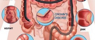
Crohn's disease, or ulcerative colitis, is classified as a chronic pathology of the human intestine with unknown causes. However, the connection of the disease with impaired immunity and the body’s aggression against its cells has been established, so treatment includes immunosuppressive drugs. Heredity also plays a role. Ulcers, adhesions, and fistulas form in different parts of the intestine. The mucous membrane swells and thickens, fibrous irreversible changes occur in it.
Symptoms and clinical picture of the disease depend on the location of the lesion. The small intestine is affected in 10-15% of cases. The symptoms are as follows: nausea, vomiting, fever, severe pain, blood in the stool, refusal to eat, general symptoms (weakness, fatigue, fever). The pain is localized in the umbilical and hypogastric region. Begins 2-4 hours after eating. The stool becomes more frequent and becomes liquid. Positive symptoms of peritoneal irritation. Treatment of the disease is long and requires patience and strict adherence to doctor’s prescriptions.
To diagnose diseases, examination of the small intestine is used using the following methods:
- X-ray contrast. To determine the structure and functional features. A barium suspension is used as a contrast.
- Endoscopic method. Allows you to visually assess the condition of the mucous membrane, sphincters, and take a biopsy for histological examination.
- Stool examination. Evaluate macroscopically, microscopically and chemical composition.
- Ultrasound of the intestine using contrast. In many ways similar to x-ray examination.
- Magnetic resonance imaging (detects diseases of a tumor nature).
- CT scan. Informative methods for detecting volumetric processes.
- A modern method using a capsule with video recording (capsule endoscopy makes it possible to check the entire intestine and identify diseases of any nature).
- Enterography (introduction of a probe and contrast into the small intestine).