Acute dilatation of the stomach in humans: what is it?
Acute dilatation of the stomach belongs to the category of uncommon diseases of this organ and the gastrointestinal tract as a whole. On the other hand, at its first signs, clinical alertness should arise, since the consequences of the described disease can be severe and lead to critical consequences.
Males get sick more often than females. But women often suffer from acute dilatation of the stomach more severely.
note
If acute dilatation of the stomach is diagnosed in childhood or adolescence, this indicates some kind of disturbance in the intrauterine development of the fetus.
Causes of acute gastric dilatation
Acute dilatation of the stomach is a polyetiological disease - this means that its occurrence is provoked by a number of factors, and they can be very different in nature.
For the purpose of systematization, all the causes of this disease are divided into groups:
- neurological;
- nutritional (food);
- infectious;
- somatic;
- tumor;
- mental;
- bad habits;
- medicinal.
The neurological reasons that provoke the occurrence of the described disorder are pathologies of the peripheral and central nervous system, due to which the nervous regulation of the stomach is disrupted. They can be inflammatory, traumatic, tumor or other in nature.
The nutritional (food) causes of the development of this disease include alternating diets with severe restrictions and overeating.
It is assumed that the bacterium Helicobacter pylori, which inhabits this organ, may be involved in the occurrence of acute dilation of the stomach. Also, this condition developed after an inflammatory lesion of the stomach with the addition of staphylococci, streptococci, E. coli and some specific pathogens (in particular, Mycobacterium tuberculosis).
Of the somatic disorders, most often the background for the development of the described pathology are:
- formation of stones in the bile duct system;
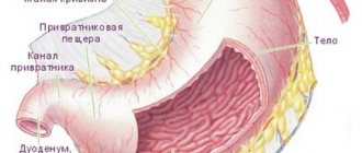
- pyloric stenosis;
- his scars
and so on.
Of the tumors, any neoplasm that becomes a barrier to the food masses evacuating from the stomach is important for acute expansion of the stomach. These can be either benign or malignant tumors.
What bad habits can provoke the development of the described pathology? This includes smoking and drinking alcoholic beverages. Nicotine constricts blood vessels (including in the wall of the stomach), due to which blood flow worsens, less nutrients (fats, proteins, carbohydrates) reach the tissues of the gastric wall, it becomes depleted and then stops contracting as usual.
note
Drug-induced causes of acute gastric dilatation most often are drugs administered to the patient to ensure medicated sleep during surgery.
Causes
Complex pathologies lead to the appearance of this disorder. These include:
- traumatic injuries of the spinal column, brain, abdominal cavity;
- infection of the biliary or urinary tract;
- urolithiasis or cholelithiasis;
- acute form of pancreatitis;
- constant feeling of hunger or severe overeating;
- ulcerative lesion or tumor of the stomach;
- intestinal obstruction;
- drinking excessive amounts of alcohol;
- childbirth;
- complex forms of infectious diseases.
In addition, the development of the disease may be the result of obstruction of the pyloric zone of the organ, which develops when the duodenum is compressed by blood vessels.
Development of pathology
Even if the root cause of acute gastric dilatation is not neurological disorders, the end result, regardless of the etiological factor, is the disruption of the nervous supply of this organ. Bioelectric impulses that move along neurons begin to move faster and more slowly - the muscular wall of the stomach reacts to such a slowdown: its tone sharply decreases, it practically loses the ability to contract.
The contents that have accumulated in the lumen of the stomach put pressure on its walls, and violations on their part lead to greater development of atony of the gastric wall. Thus, a kind of vicious circle is formed.
note
In acute dilatation of the stomach, the disorder is that the gastric wall loses its ability to absorb liquid, while its secretory (excretory) function is preserved.
With the disease described, not only the stomach suffers. It, being overfilled with liquid contents, shifts, which causes the organs adjacent to it to shift - the duodenum, small intestine, jejunum and loops of the large intestine. They are attached to the abdominal wall by the mesentery, which becomes stretched with such displacement. The mesentery contains blood vessels and nerve structures of the small and large intestines, so if it is damaged, then these elements suffer.
The following types of acute gastric dilatation have been identified:
- by degree of increase - initial, medium, pronounced;
- according to the presence of consequences - complicated, uncomplicated;
- by drawing neighboring organs into the pathological process;
- by type of expansion - primary (initiated by a large amount of food in the lumen of the stomach), secondary (associated with damage to the nervous apparatus of the gastric wall).
Reduction procedure
If a person notices regular overeating, but feels slight discomfort due to the slow absorption of consumed products, drastic measures are not required. It is enough to carry out a light massage of the abdominal muscles with the palm of your hand. To do this, you need to massage your stomach with smooth clockwise strokes. A one-time dose of enzymes is allowed to improve digestive function. These measures will speed up the process of emptying the stomach, prevent prolonged stretching of the walls and advanced hypertrophy with consequences.
If a slight change in organ volume is detected, gentle treatment with a diet and dietary adjustments is permissible. The course of diet therapy is 4-6 months. Objectives: split meals (up to 6 times a day) in small portions (up to 300 ml in total with solid food and liquid. During the treatment period, excessive consumption of fats should be avoided. In case of severe distension of the stomach tissues with severe hypertrophy, two ways to solve the problem are recommended:
- operation;
- activation of the natural process of tissue contraction to its previous state.
Why one or another technique is chosen depends on the severity of the deformity, the condition of other organs and systems of the body, the duration of the treatment course, and psychological aspects.
Return to contents
Symptoms of acute gastric dilatation
Acute gastric dilatation is an acute condition. The clinical picture develops quite quickly, the disorders progress before our eyes. There is a deterioration in the patient's condition literally over the course of one hour.
The clinical picture is represented by the following signs:
- local;
- are common.
The main local symptoms of acute gastric dilatation are:
- pain;
- hiccups;
- dyspeptic disorders.
The characteristics of pain in this pathology will be as follows:
- by localization - in the epigastric region;
- by distribution - pain can spread to neighboring locations, and as the pathology progresses - to the entire abdominal cavity, while the patient seems to have pain throughout his entire abdomen;
- by nature – pressing, bursting, spasmodic, sharp;
- in terms of severity - medium intensity or strong, increasing with the progression of pathology and an increase in the amount of stagnant contents in the stomach;
- by occurrence - they appear when contents accumulate in the stomach, occupying about a fifth of this organ; with a progressive increase in its quantity, they increase.
note
With acute dilatation of the stomach, abdominal pain is caused not only by stretching of the stomach, but also by uncontrollable vomiting, due to which the anterior abdominal wall becomes tense.
Hiccups with acute dilatation of the stomach occur reflexively.
Dyspeptic disorders that occur with stromal dilatation of the stomach are:
- nausea;
- vomit;
- worsening of gas discharge;
- violation of the act of defecation.
Nausea with acute dilatation of the stomach is constantly observed, accompanied by the urge to vomit.
Vomiting is the most important symptom of the described disease. Its characteristics are as follows:
by occurrence - as a rule, it appears at the time of an acute painful attack from the stomach;- in terms of frequency – very frequent, the patient does not have time to literally catch his breath after one attack of vomiting before the next one begins;
- by nature - gushing, with vomit literally pouring out from the gastrointestinal tract;
- in terms of the amount of vomit - on average from five to ten liters per day;
- by the nature of the vomit - they are stagnant stomach contents. In particularly severe forms of vomiting, it may contain impurities of bile and coffee grounds. The second signals a violation of the integrity of the gastrointestinal tract vessels and bleeding - the blood mixes with the gastric contents, forming a specific brown substance;
- According to the patient’s subjective feelings, vomiting does not bring relief, and if it is prolonged, it leads to exhaustion of the patient’s strength, who is no longer able to tolerate convulsive contractions of the gastric wall.
note
It is characteristic that after some time the number of vomiting “acts” and the volume of vomit decrease. This is explained by the patient's dehydration, so a decrease in the intensity of vomiting is an alarming diagnostic sign.
Since the intestines are not affected, gases continue to pass, but due to the triggering of the reflex mechanism of the intestinal response, it is worse.
A patient with acute gastric dilatation has irregular diarrhea.
General symptoms of acute gastric dilatation occur due to the body losing a large amount of fluid. The following signs are observed:
- general weakness;
- feeling overwhelmed;
- severe impairment of ability to work – mental and physical;
- lethargy;
- “flies” before the eyes;
- decrease in the amount of urine excreted per day;
- decrease in body temperature.
Symptoms
The primary expansion of the organ is accompanied by the sudden onset of pain, which affects the entire abdomen and progresses rapidly. In this case, the passage of gases is disrupted. Gradually, severe vomiting appears - first, the acidic contents of the stomach are removed, and then bile. Subsequently, venous stasis and bleeding cause the vomit to turn brown.
First, the person experiences increased arousal and tries to take a comfortable position. Typically, relief occurs in the knee-elbow position or lying on the right side with legs raised.
Then the development of collapse is observed as a result of severe dehydration, which provokes oliguria and anuria. Due to increased loss of electrolytes, seizures may occur.
The entire abdomen is greatly distended, but the iliac zones remain normal. This is a specific manifestation of gastric dilatation. As a rule, the border of the enlarged organ can be clearly visualized.
There are no signs of irritation of the peritoneum, however, when palpated, the abdomen is quite tight. When tapping, you can detect the sound of splashing.
Diagnostics
The diagnosis of acute gastric dilatation is made through a comprehensive examination - the patient’s complaints, details of the anamnesis (history) of pathology, and the results of additional examination methods (physical, instrumental, laboratory) are taken into account.
From the anamnesis, the following is revealed:
- what does the patient associate with the clinical picture that has developed;
- how often does vomiting occur?
- whether the patient previously suffered from any stomach pathologies.
Physiological examination reveals the following abnormalities:
- upon general examination, the patient’s general condition is of moderate severity, and with the progression of the disease (in particular, uncontrollable vomiting), the condition is close to severe. The skin and visible mucous membranes are pale, the skin is dry due to dehydration due to vomiting;
- upon local examination, the tongue is dry, covered with a white coating, and an unpleasant putrid or fermentative odor may be felt from the mouth. In thin patients, there is a protrusion in the epigastric region due to an enlarged stomach. With flatulence, bloating is visualized;
- during palpation (palpation) – pain in the epigastric region and fluctuation (fluctuations of the fluid during pushing movements with the examiner’s fingers) are noted. If flatulence occurs, its presence is also confirmed by palpation;
- during percussion (tapping) - at the site of projection of the overfilled stomach onto the anterior abdominal wall, a dull sound is noted when tapping. In the lower sections, against the background of flatulence, the sound will be similar to that which occurs when tapping on a hollow object;
- during auscultation (listening with a phonendoscope), a splashing noise is noted. Over the lower abdomen, peristaltic bowel sounds are weakened.
Instrumental research methods that are used in the diagnosis of the described disease are:
- plain radiography of the abdominal organs. Despite the fact that this diagnostic method is generally less informative than other methods, in this case it plays an important role: X-ray images show a distended stomach, while neighboring organs are displaced. The increase in the volume of the stomach can reach such an extent that its lower edge reaches the pelvic region;
- Contrast radiography - the patient ingests a portion of a suspension of barium sulfate (a contrast agent), after which a series of X-ray images are taken. In the photographs, the stomach is presented as a shapeless bag overflowing with a contrast agent. Also, due to their serial nature, the images allow one to estimate the rate of evacuation (release) of the contrast agent from the stomach - with its acute expansion, they are critically slowed down or evacuation does not occur at all;
- fibrogastroduodenoscopy (FGDS) - a fibrogastroscope (a type of endoscopic equipment) is inserted into the stomach through the esophagus, examined from the inside, and the size of the organ is assessed using indirect signs. It is often difficult to evaluate what is seen during FGDS, since the stomach is filled with mucus, gastric juice and fermented food masses that are not evacuated into the duodenum due to atony of the gastric wall;
- computed tomography (CT) - when carrying out this research method, layer-by-layer images-sections of the tissue of the gastric wall are taken, which allow one to evaluate its structure. Thanks to this, the information content of the method is very high and allows you to identify violations;
- multislice computed tomography (MSCT) is a type of computed tomography, but more advanced;
- Magnetic resonance imaging (MRI) - during this procedure, layer-by-layer images of the gastric wall are also obtained. But MRI, in comparison with CT and MSCT, is more informative when studying soft tissues.
Laboratory research methods used in the diagnosis of acute gastric dilatation are:
general blood test - the most characteristic sign is a violation of hematocrit (the ratio of formed elements and the liquid part of the blood). With prolonged pathology, due to the addition of an infectious agent, an increase in the number of leukocytes (leukocytosis) and ESR is observed;- biochemical blood test - there is a decrease in the amount of sodium, potassium, chlorine, and an increase in the amount of nitrogen compounds. The first occurs due to profuse vomiting, the second due to the fact that stagnant gastric contents begin to ferment, fermentation products are absorbed into the blood, reach the liver and kidneys and negatively affect their function in removing nitrogenous products of tissue metabolism;
- microscopy of gastric contents - it reveals undigested food elements, and at later stages of pathology development - an attached infectious agent.
Differential diagnosis
Differential (distinctive) diagnosis of acute gastric dilatation is most often carried out with diseases and pathological conditions such as:
- gastritis is an inflammatory lesion of the gastric mucosa. Differential diagnosis should be carried out with the acute form of this pathology;
- duodenitis - inflammation of the duodenal mucosa;
- gastric bezoar is a foreign body formed from undigested elements that were swallowed accidentally or purposefully;
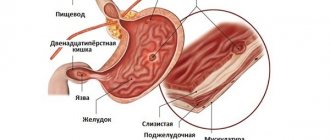
- Gastric ulcer is the formation of deep destruction of the gastric wall. Differential diagnosis should be carried out with this pathology in the acute stage;
- perforated ulcer - the occurrence of a through defect in the wall of the stomach at the site of ulceration, which is accompanied by the release of gastric contents into the abdominal cavity;
- penetrating ulcer – spread of the ulcerative process from the stomach wall to neighboring organs;
- pancreatitis - an inflammatory process in the parenchyma (working tissue) of the pancreas;
- pancreatic necrosis – necrosis of the pancreatic parenchyma, which occurs when simultaneously ingesting large amounts of fatty foods and alcoholic beverages;
- intestinal obstruction - a violation of the passage of food masses through the intestinal lumen;
- food toxicoinfection – poisoning by food products containing pathogenic microflora;
- dysentery - acute infection of the intestines by Shigella, which is characterized by profuse repeated diarrhea;
- peritonitis is an inflammatory lesion of the peritoneum (the connective tissue film that lines the inside of the abdominal wall and covers its organs).
Diagnostic methods
CT can be used to diagnose pathology.
The examination is carried out by palpation by a gastroenterologist or surgeon. Diagnosis is made using fluoroscopy. Pictures are taken using contrast after taking a special liquid. An enlarged stomach is an indication for the use of conservative therapy. During gastroscopy, the mucous membrane and walls of the stomach are examined. Diagnostics is carried out using a probe with a camera, displaying an image on the screen. A sample is taken for histological analysis. Computed tomography is painless and can be performed without the use of contrast. Its advantage is the ability to view in several projections.
Complications
The main consequences of acute gastric dilatation are:
violation of the motor function of the organ - its contents are not evacuated into the duodenum;- indigestion;
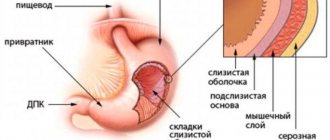
- perforation of the gastric wall - the formation of its through defect. The gastric wall itself is a strong elastic structure, but if the patient suffered from a gastric ulcer, then due to the high pressure of the contents, even greater thinning of the wall at the site of the ulcer and the formation of a through defect are possible;
- gastrointestinal bleeding.
Suturing the stomach
Gastric suturing surgeries are popular among obese people. The simplest one: a narrowing ring is placed on it and divided into two unequal sections with a narrow passage between them. Because of this, food leaves the upper chamber more slowly, and the person feels full.
It is also practiced to suture the stomach to create a pouch from the upper part, which is directly connected to the small intestine. This is not cutting off the “stretched part”, but reducing the stomach by dividing it into two chambers, after which only one “working” chamber remains. As a result, satiety occurs with a small amount of food.
Sometimes a part is cut off, but this is not an “extra” part of the stretched walls of the stomach, and the ordinary stomach is made smaller.
This limits both food intake and the amount of calories and nutrients.
The small pouch that remains is connected directly to the terminal segment of the small intestine, bypassing the upper part of the small intestine entirely. A common channel remains in which bile and pancreatic digestive juices mix before entering the large intestine. A person loses weight because most of the calories and nutrients are sent to the colon, where they are not absorbed.
Treatment of acute gastric dilatation
Treatment of the described pathology can be conservative and surgical.
Acute gastric dilatation refers to acute conditions - that is, those that require immediate intervention. First, medical care is provided aimed at improving the patient’s condition, then he is taken to the clinic, where he will undergo a comprehensive examination and treatment.
Important
In case of acute dilatation of the stomach, a nasogastric tube is inserted to remove excess contents from the stomach. In this case, it is strictly forbidden to stimulate the patient to vomit.
The main appointments in the clinic are:
- bed rest;
- hunger;
- placement of a nasogastric tube - it remains in the stomach for 24-76 hours, sometimes up to 5-6 days;
- drug therapy.
The duration of bed rest is determined by the general condition of the patient.
The contents of the stomach are not only released through the tube - it must be actively sucked out.
Medication prescriptions are as follows:
- infusion therapy;
- general strengthening appointments.
The purpose of infusion therapy is to restore the amount of fluid in the body that was lost during attacks of profuse (uncontrollable) vomiting. Saline solutions, electrolytes, glucose, blood serum, and so on are injected intravenously.
General restorative therapy means:
- parenteral nutrition - intravenous administration of protein drugs, since the patient cannot eat food in the usual way;
- vitamin therapy - the use of injectable drugs.
Surgical treatment is carried out for certain conditions that could provoke acute dilatation of the stomach. So, in case of violations on the part of the mesentery, its partial excision is performed. In some cases, it is necessary to form a communication between the stomach and the anterior abdominal wall - this will ensure gastric emptying. During surgical treatment, drug therapy is carried out in full.
Treatment methods
The choice of treatment tactics is carried out on an individual basis. For treatment to be effective, it is necessary to take into account the factors that led to the expansion of the stomach.
If the pathology is not accompanied by a strong narrowing of the exit part of the organ, conservative therapy is used. First, the stomach is emptied. Then, to relieve pressure and relieve pressure, the organ is washed. Normalization of protein and carbohydrate metabolism is of no small importance. This is achieved through parenteral nutrition and infusion therapy.
With such a diagnosis, it is imperative to restore the functioning of organs and tissues. For this purpose, a bilateral perirenal block is performed. Drugs are also prescribed to normalize gastric tone. These include drugs such as prozerin, acyclidine.
If there is a strong drop in pressure, polyglucin or polyvinol is administered intravenously. To detoxify the body, treatment is carried out with neodez and hemodez. Be sure to use ascorbic acid and B vitamins.
If conservative therapy does not produce the desired results, surgical intervention becomes necessary. The operation is performed to eliminate the cause of the disease. In this case, it is necessary to decompress the organ and establish enteral nutrition.
To empty the organ and improve the person’s condition, a gastrostomy tube is placed. With this diagnosis, eating through a tube is contraindicated. It is also prohibited to use antispasmodics or narcotic analgesics. Such drugs provoke a worsening of atony.
If the stomach ruptures and peritonitis occurs, urgent surgery is performed. In this case, the integrity of the organ is restored, the abdominal cavity is cleansed and drained.
If the pathology of the stomach is accompanied by a narrowing of its outlet part, resection of the organ is performed. If measures are not taken promptly, this disease can be fatal.
Prevention
Measures to prevent acute gastric dilatation are:
prevention, identification and treatment of neurological disorders, against the background of which the innervation of the stomach may suffer with the subsequent development of the described condition;- adherence to the principles of proper nutrition, avoiding strict diets and overeating;
- neutralization of the bacterium Helicobacter pylori. For this purpose, courses of antibacterial therapy are carried out;
- prevention, diagnosis and relief of somatic pathologies that can cause acute dilatation of the stomach - cholelithiasis, pyloric stenosis, its scars, and so on;
- prevention, detection and elimination of tumor pathologies of the stomach;
- rejection of bad habits;
- correct administration of anesthesia.
Risk of complications
If necessary, doctors can resort to bandaging the organ.
If conservative treatment is ineffective, surgical intervention is performed. With gastrostomy, a gastric stoma is created. Drainage is in progress. Techniques without tissue removal are used. For example, bypass surgery, bandaging or sewing in a silicone ball. Complications associated with surgery have an adverse effect on the human body.
The most threatening complication of the disease is gastric rupture. Develops against the background of shock and peritonitis. Most cases are fatal.
Forecast
The prognosis for acute gastric dilatation is generally favorable. With the help of competent prescriptions, you can stop the causes of this pathology and its consequences.
At the same time, complications of this disease occur quite often: bleeding of varying severity and perforation of the gastric wall are observed, according to some data, in every fifth patient with this diagnosis.
The prognosis worsens with:
- complications;
- late relieved dehydration.
Kovtonyuk Oksana Vladimirovna, medical observer, surgeon, consultant doctor
1, total, today
( 59 votes, average: 4.88 out of 5)
Stomach pain near the navel: what to do, possible causes
Stomach pain: causes, characteristics, treatment
Related Posts
Disease provocateurs
The main cause of the problem is obesity, but diseases of the digestive system can also provoke an enlarged stomach.
In most cases, the digestive organ is enlarged due to obesity. But the reason why the stomach can increase in size lies in the influence of other provoking factors, such as diseases of the gastrointestinal tract and other organs and systems. These include:
- Pyloric narrowing. In this case, hypertrophy is accompanied by a sour taste in the mouth, a persistent feeling of fullness in the stomach, and frequent vomiting.
- Cancer. The disease is manifested by constant heaviness in the stomach, the presence of blood in the stool, severe weakness, and decreased appetite.
- Hernia of the stomach or esophagus. In this case, gastric hypertrophy is accompanied by pain when eating and when changing body position.
- Gastric obstruction. With hypertrophy, in this case, food stagnates in the stomach, which provokes the formation of polyps and tumors, which means an increase in the organ.
- Hyperplastic gastropathy. The disease is accompanied by an increase and thickening of folds in the epithelial mucosa of the stomach, which leads to an increase in the size of the organ. The process causes delayed digestion, bloating, pain in the left hypochondrium, and heaviness.
- Ménétrier's disease, characterized by the formation of polyposis in the stomach, which increases its size. Due to polyps, the walls of the organ expand, accompanied by frequent pain in the left abdominal region, weight loss, nausea, diarrhea, and bleeding.
Return to contents
