Diagnostic interview and examination
At the first meeting with the patient, the doctor is interested in complaints and conducts a general examination of the patient. During the conversation, the doctor learns the characteristics of pain, the nature of dyspepsia, the frequency and intensity of clinical symptoms. The following symptoms have the greatest diagnostic value in diagnosing gland diseases:
- Pain in the upper abdomen, mainly in the epigastric and subcostal areas. The pain is often girdling in nature and occurs after eating a large fatty meal. The heaviness and pain in the abdomen does not go away for a long time.
- Painful sensations radiate to the left shoulder blade and lower back, which forces the person to take a forced position to relieve the condition.
- A characteristic symptom of pancreatic disease is vomiting and nausea after eating fatty foods. Bile may be present in the vomit. Vomiting does not relieve the pain symptom.
- In diseases of the gland, due to insufficient digestion of fats and lipids, steatorrhea occurs - frequent liquid or pasty yellow stools mixed with fats. Steatorrhea is a pathognomonic symptom in the diagnosis of pancreatitis, tumor and organ cancer
- Patients are periodically bothered by bloating, fever, symptoms of intoxication, and icteric discoloration of the skin, which also indicates pancreatic disease.
Important information! Due to enzyme deficiency, some patients note unmotivated weight loss, which may also speak in favor of gland pathology. If this symptom appears, you should immediately consult a doctor, since in the vast majority of cases, weight loss is a sign of the development of a cancerous tumor in the body.
An external examination reveals jaundice and dry skin. On palpation, pain is detected in the projection areas of the pancreas, but it is not possible to completely examine and palpate the organ due to its deep location.
Laboratory diagnostic methods
The second stage of the diagnostic search is laboratory tests. For this purpose, clinical and biochemical blood tests, clinical and biochemical urine tests, stool microscopy (coproscopy), and functional tests to detect digestive enzyme deficiency are prescribed.
Clinical blood test
With inflammation, the hemogram reveals leukocytosis and accelerated ESR. The addition of a purulent infection is characterized by a shift in the leukocyte formula. With cancer, the amount of hemoglobin, red blood cells, and platelets in the blood decreases.
Blood chemistry
- First of all, the amount of amylase (pancreatic enzyme) is assessed; with pathology of the organ, amylase in the blood increases tens of times.
- Next, if possible, the amount of more specific enzymes is assessed: lipase, elastase, the amount of which in the blood also increases.
- Inflammation of the organ is indicated by dysproteinemia (violation of the ratio of protein fractions) and the appearance of C-reactive protein.
- Secondary damage to the pancreas due to diseases of the biliary and hepatolienal systems is indicated by an increase in bilirubin, transaminases (AST, ALT), alkaline phosphatase, GamaGTP.
- With cancer and tumors there are no specific changes in the blood. The neoplasm may be accompanied by any of the above symptoms.
| Biochemical indicator | Norm | Changes in gland pathology |
| Protein | 65-85 g/l | Dysproteinemia: an increase in total protein mainly due to the globulin fraction. |
| Fasting glucose | 3.3-5.5 mmol/l | Increased due to parenchymal atrophy and decreased insulin production |
| Transaminases (AST, ALT) | AST – up to 40 U/l ALT – up to 45 U/l | Promotion |
| Alkaline phosphatase | Up to 145 U/l | Increased with cholestasis |
| C-reactive protein | Absent | Appears |
| Amylase | Up to 50 U/l | Tenfold increase |
| Elastase, lipase | Up to 5 mg/l | Promoted |
Biochemical analysis of urine for diastase
The main method for diagnosing acute and chronic pancreatitis in the acute phase. At the same time, a high content of diastase (alpha-amylase) is detected in the urine - a specific sign of pancreatitis.
Stool examination
Microscopy of feces is carried out to diagnose digestive enzyme deficiency. The test is considered positive when undigested lipids, fats, and muscle fibers are detected. This symptom is characteristic of both inflammation and gland cancer. If possible, the amount of pancreatic elastase and lipase, which are also detected in large quantities, is determined in the stool.
Coprography for pancreatic dysfunction
The primary sign of pancreatic dysfunction is the release of increased amounts of fat in the stool. According to research by the World Health Organization, the normal level of fat excretion through feces is no more than 7 g when consuming 100 g of fatty foods. An increase in this indicator indicates that the gland does not produce a sufficient level of enzymes to break down fats, as a result of which they are excreted undigested.
Examination of the pancreas using this test necessarily involves a strict diet for at least several days. It was developed according to Schmidt's conditions:
- daily protein - 105 g;
- daily fat intake - 135 g;
- Approximately 180 g of carbohydrates should enter the body.
Important information: How to check the pancreas and what tests to take
Such nutrition to check the pancreas gives the most complete picture of further bowel movements. It is as balanced as possible (the size can be changed proportionally on the doctor’s recommendation to meet the needs of the body), and with the correct functioning of the gastrointestinal tract, deviations in stool are impossible with such a diet.
Several factors can affect the clarity of the tests a patient must take. Consuming alcohol and fatty foods has a negative effect on the results. All this makes the enzymes less active. It is prohibited to take drugs that are enzymatic before donating stool. They can compensate for the lack of their own substance in the body and hide the symptom from the doctor.
If muscle tissue is detected, poorly digested and abundantly excreted in the feces, one can judge about diseases of other parts of the gastrointestinal tract - the intestines or stomach. It is important to follow all the rules for conducting tests when diagnosing, otherwise the data obtained will not correspond to reality. Delayed diagnosis also means delayed therapy, increasing the risk of complications.
Functional tests
Most informative for severe enzyme deficiency. Currently, they have limited use, since more effective X-ray techniques for examining patients have appeared.
For diseases of the pancreas, the Lund test is used (probing of the duodenum after a test breakfast, followed by aspiration of the contents and its biochemical examination), a radioisotope test (to detect steatorrhea), a glucose tolerance test (if a decrease in insulin production is suspected), a pancreatolauric test, etc. Explanation of the results tests are carried out by a doctor, a diagnosis is made only when the data is confirmed by clinical symptoms.
Important! If cancer or a benign tumor is suspected, the blood must be tested for tumor markers.
General rules for preparing for analysis
To determine the disease, tests are taken, especially if pancreatitis is suspected. How to examine the pancreas and get the correct tests after diagnosing the body? This is a sensitive issue, since errors in collecting the required biomaterial will lead to some deviations and the prescription of incorrect treatment.
For the diagnostic procedure itself, general requirements have been developed, which include:
- Pancreatic tests are taken on an empty stomach, in the morning. In 1-2 days, stop eating salty, spicy, fatty foods, try to give up bad habits and alcohol, stop drinking carbonated water and legumes.
- To draw blood, stop smoking at least two hours in advance.
- If the patient has constipation, then it is necessary to cleanse the intestines with an enema and take enterosorbents (activated carbon and many others). After all, the accumulation of digested food has a toxic environment and will spoil the complete picture of the diagnosis of the body.
- All containers for test material are sterile, hands are washed with soap.
- For females, before donating urine, perform hygiene procedures with the genitals.
- When taking a general urine test, the middle part of the portion is taken.
The pancreas and its diagnosis require compliance with the general rules for collecting material for diagnosis. The correctness of the results obtained determines the clinical picture of treatment for pancreatitis or other complications of this disease.
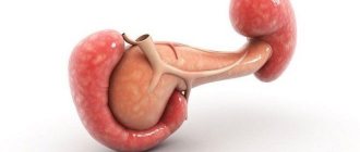
location of the pancreas
In addition to diagnosing your health condition, there are symptoms that, together with the test data obtained, will confirm the disease pancreatitis:
- diarrhea;
- girdle pain;
- gagging;
- severe weakness in the body;
- sudden appearance of pain in the solar plexus and side of the stomach.
If such symptoms appear, urgently visit a medical facility and get tested for the pancreas and side diseases of pancreatitis. And also try to determine the disease yourself. It happens that it is not possible to visit a medical facility, therefore, based on the existing signs, you can understand at home that the pancreas hurts.
manifestation of the acute phase after alcohol and fatty foods
The acute phase of the disease mainly manifests itself after heavy consumption of alcohol or fatty foods, which gives impetus to the inflammatory process. In this case, a sharp girdling pain occurs, which extends to the back and intensifies when lying down. The pain will be dulled by lying on your side and tucking your knees under your stomach. In the acute phase of exacerbation, analgesics may not bring positive results.
Also, the condition of the affected person is aggravated by vomiting, bloating, and yellowed sclera of the eyes. In such a situation, self-medication is dangerous to health and requires urgent diagnosis. When you visit a doctor, he prescribes tests to get a complete picture of pancreatic disease, which will make it possible to correctly prescribe treatment.
In the chronic form of the disease, the symptoms differ slightly from the acute form of pancreatitis:
- gradual weight loss;
- periodic pain in the right and left hypochondrium;
- diarrhea with a strong odor and light-colored stool;
- vomiting with constant nausea;
- dry mouth;
- thirst;
- feeling of uncontrollable and constant hunger.
Without a medical education, a person can make an inaccurate diagnosis for himself. This will do you no good, so first of all, find a way to undergo diagnostic tests to identify damage to the pancreas.
tests for pancreatitis and inflammation of the pancreas
What tests are there for pancreatitis and inflammation of the pancreas:
- General blood analysis.
- Biochemical blood test.
- Stool analysis.
Laboratory tests will help establish a diagnosis and determine the inflammatory process in the pancreas. The most important thing about them is the detection of the amount of enzymes in the blood. On the first day of exacerbation, they look at pancreatic amylase, on the second - the volumetric content of lipase and elastase.
Instrumental diagnostic methods
Confirmation of the diagnosis is impossible without instrumental methods. At the present stage of development of medicine, X-ray, ultrasound and fiber optic diagnostic methods are used.
X-ray studies
- Plain radiography of the abdominal cavity. Used for differential diagnosis of abdominal pain syndrome. Indirect signs of damage to the pancreas are stones and compactions in the gallbladder and bile ducts.
- Endoscopic retrograde cholangiopancreatography (ERCP). The method is also effective for secondary biliary-dependent pancreatitis due to congestion in the bile ducts, stones in the gall bladder, and cicatricial narrowing of the excretory ducts.
- CT scan. Helps diagnose complicated pancreatitis (cysts, pseudocysts, calcifications, atrophic and necrotic areas of the organ). Widely used for large tumors: benign gland tumors, cancer, cancer metastases from neighboring organs. With these pathologies in the images, the contours of the gland are uneven, the size is increased, and a voluminous neoplasm is detected in the area of one or two lobes.
Ultrasonography
Ultrasound of the abdominal organs and, in particular, the pancreas is the gold standard for diagnosing primary and cholangiogenic pancreatitis, fatty and connective tissue degeneration of the parenchyma, and pancreatic cancer. In conclusion, the doctor gives an accurate description of the structure of the organ, the severity of diffuse changes, their nature and prevalence.
- With stones in the gall bladder or excretory ducts, dense stones of varying sizes and densities are visualized.
- In acute and chronic pancreatitis, diffuse changes in the parenchyma are detected in all parts of the organ in combination with edema of the capsule and interlobular spaces.
- With cancer, the size of the organ is increased, the echogenicity of the structures is not uniform. The monitor clearly shows the boundary between healthy parenchyma and cancerous tissue. Based on the density of the tumor, one can judge the origin of the tumor.
Important information! If cancer is suspected, a biopsy of pancreatic tissue is performed, followed by microscopy of the structures. In case of cancer, a violation of the cytoarchitectonics of the biopsy specimen is visualized in the specimen: in the parenchyma there are multiple atypical cells with their irregular location.
Esophagogastroduodenoscopy
Another method for diagnosing pathology of the pancreas and biliary tract. The method allows you to identify cicatricial narrowing or blockage of the excretory duct with stones in biliary-dependent pancreatitis, as well as visualize changes in the pancreaticoduodenal zone, which indicates primary pancreatitis or organ cancer.
Thus, the diagnosis of pancreatic pathology is a whole complex of diagnostic studies that are carried out on the patient immediately upon admission to the clinic. All tests are prescribed by a gastroenterologist or therapist after a thorough examination and interview of the patient. The same doctor prescribes treatment.
A timely diagnosis allows you to quickly determine the direction of treatment (refer the patient to a surgical or therapeutic hospital), prescribe adequate etiotropic and symptomatic therapy, and improve the prognosis of the disease.
Biopsy for pancreas
Taking soft tissue is provided in case of suspected neoplasm. The specialist who conducts these tests using ultrasound or an X-ray machine finds the problem area, and then takes a piece of tissue from a certain area of the pancreas. A similar study is prescribed for:
- sudden weight loss;
- the appearance of cancer antigens in the blood;
- intoxication of the body for no apparent reason;
- the appearance of constant pain in the pancreas area;
- frequent bloating, digestive and metabolic disorders.
This is a second-stage diagnostic method, that is, it must be preceded by another. Before the biopsy you must:
- detect a suspicious area using palpation or penetrating radiation;
- differentiate the contents of this area as a probable tumor.
Without suspicion of neoplasms, this procedure is not performed due to the high cost of the operation and its pain.
Punctures are done in several ways: endoscopy, through a syringe without breaking the skin, or surgically. A biopsy, even with a syringe, must be performed under anesthesia, since penetration of a foreign body through several layers of biopsy tissue is fraught with severe discomfort.
According to medical rules, it is prohibited to cause severe pain to the client.
Patients are interested in whether a pancreatic biopsy is performed and the cost of the procedure. Although the study is one of the most expensive, you can afford it: 1,300 rubles per puncture are charged in clinics in the capital.
Important information: Herbs for the treatment of pancreas


