- November 13, 2018
- Gastroenterology
- Svetlana Karetnikova
Gastritis is a widespread disease. The diet of a modern person often consists of snacks, which negatively affects the condition of the stomach. Therefore, the number of patients with this disease is growing every day. But stomach pain is not always associated with gastritis. To identify it, you should undergo a thorough examination, which includes several stages. You can learn about examination methods and which doctor to contact for gastritis in this article.
What does gastritis mean?
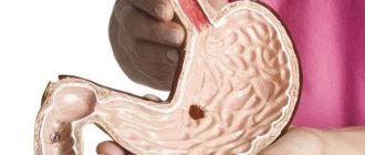
Gastritis is a disease that is accompanied by acute or chronic inflammatory-dystrophic changes in the gastric mucosa. The disease may differ in its origin, symptoms and course. The main symptoms are pain in the epigastric region, dyspepsia, heartburn and nausea. The concept of gastritis includes inflammatory and dystrophic processes in the gastric mucosa; they can spread to the duodenum. In both cases, the disease can develop into an ulcer and cause internal bleeding. Therefore, gastritis should be treated as soon as possible.
The disease can be caused by either increased or decreased acidity. The gastric mucosa suffers the most, which leads to a decrease in the production of protective mucus and some components of gastric juice. Due to problems with functioning, problems appear with digestion of food. The disease is widespread in different segments of the population and age categories. But the acute form is more common in people over 60 years of age. According to the diagnostic results, chronic gastritis was detected in 80% of the total number of all stomach diseases. The main causative agent of the disease is the bacteria H. pylori, which is found in 75-80% of the population.
Preparing for diagnosis
Before conducting laboratory and instrumental studies, it is necessary to undergo training to obtain reliable results. Patients must follow the general rules:
- Tests are taken on an empty stomach 12 hours before the material is collected;
- a few days before the examination, it is necessary to exclude harmful foods from the diet: spicy, salty, fried, smoked foods;
- giving up bad habits (alcohol, tobacco products) 2-3 days before diagnosis;
- Before donating blood, you must avoid physical activity;
- You should refrain from taking medications at least 7 days before the scheduled examination;
- Before donating feces, you should not eat foods that change the color and texture of biological material: beets, grapes, cabbage, carrots, radishes.
Test results can be affected by genetic characteristics, emotional tension, stress, physical fatigue, and recent illnesses. Therefore, these factors must be avoided before undergoing the study. Blood tests for gastritis, urine, and stool can be taken at a public clinic or private laboratories.
Diagnostics is a mandatory stage in identifying the occurrence of inflammatory processes in the mucous membrane of the digestive organ and prescribing treatment methods. Tests for gastritis are a necessary procedure to identify the causes of the development of pathology. Diagnosis involves various research methods depending on the nature of the symptomatic signs of the disease. Timely medical examination will prevent the progression of chronic, acute gastritis.
We recommend: How to get rid of bad breath with gastritis?
Types and classification of gastritis
The disease is divided into two types:
- acute gastritis – characterized by a one-time exposure to irritants, manifests itself once;
- chronic gastritis - the disease systematically recurs.
Acute gastritis, in turn, is divided into four types:
- Catarrhal. It appears under the influence of food poisoning or poor nutrition.
- Fibrinous. Occurs under the influence of sublimate poisons or acids, as well as in complex infectious diseases.
- Corrosive. Appears after alkalis, heavy metal salts, and concentrated acids enter the stomach.
- Phlegmonous. It occurs as a result of complications or injuries after ulcers or stomach cancer and some infectious diseases.
In accordance with the classification according to the pathogenetic mechanism, when diagnosing gastritis (including chronic), it can be classified as one of the following types:
- type A, or autoimmune, is gastritis that appears under the influence of antibodies to the lining cells of the stomach;
- type B, or bacterial gastritis, occurs due to the harmful effects of the bacteria H. pylori;
- type C, or chemical gastritis, it appears under the influence of the reflux of lysolecithin and bile into the stomach.
Mixed AS or AB and alcoholic, drug-induced and other types of gastritis are also distinguished, depending on the origin.
Diagnosis of chronic gastritis
If you observe the above changes in general health in yourself or your loved ones, you should immediately seek help from a doctor. Usually, this is a general practitioner who, after examining and talking with the patient, will decide what additional examinations and highly professional consultations are needed.
Despite the apparent simplicity and frequency of occurrence of gastritis, including chronic gastritis, the diagnosis of this disease is a process that requires time, special tests and research methods. To successfully get rid of the pathology or significantly improve the condition and achieve long-term remission, it is necessary to conduct a full examination to accurately establish the etiology (cause) of inflammation of the gastric wall.
How to identify gastritis?
The disease usually appears unexpectedly. Pain in the upper abdomen begins sharply. They usually occur on an empty stomach and are cramp-like in nature. The pain is accompanied by nausea or vomiting, as well as heartburn, “sour” belching, a feeling of pressure and fullness in the epigastric region, as well as bloating. In the chronic form of gastritis, the patient often experiences diarrhea or constipation. The disease affects the general well-being of a person. He feels lethargic and drowsy, fatigue increases and irritability occurs. Gastritis affects the tone of the body and can affect the cardiac system, causing numbness in the upper and lower extremities and a burning sensation in the mouth and tongue.
The disease can occur without obvious signs, especially in a chronic form. But, as a rule, the first symptoms of gastritis are sharp pain in the epigastric region, which lasts a short time. The patient also periodically experiences heartburn and nausea. He has problems with bowel movements, which may alternate between constipation and diarrhea. The pain is often accompanied by vomiting, bleeding and dizziness.
After the first signs appear, diarrhea may occur throughout the day. If this happens, you need to completely stop eating food and urgently go to the hospital where your stomach will be washed out. If the symptoms are accompanied by bleeding, the patient is injected with drugs that reduce the synthesis of hydrochloric acid. In this case, medications are prescribed to promote the healing of erosions.
Differential diagnosis of gastritis
To establish an accurate diagnosis, the doctor talks with the patient and listens to his complaints. Next, he conducts a physical diagnosis of gastritis using palpation of the stomach area. If a person experiences a feeling of discomfort, this may indicate the presence of an inflammatory process, and acute pain may indicate the development of gastritis. The specialist must also examine the patient’s oral cavity. The presence of a yellow tint is a sign of acute corrosive gastritis. An increased content of sulfuric acid is indicated by black spots in the oral cavity, white spots indicate hydrochloric acid. The presence of a grayish-brown film is a sign of alkali poisoning.
During the initial examination, gastritis can manifest itself in the form of elevated temperature, increased muscle tone in the stomach area, as well as the person’s general well-being. But in order to confirm the diagnosis, further examination must be performed.
Clinical examination
Initially, a clinical examination of the patient takes place, during which the doctor examines the condition of the skin, palpates the abdomen where the stomach is located: if the patient has gastritis, the pain worsens.
Clinical examination gives only a guess about the disease. To find out the extent of damage to the stomach and its nature, the use of instrumental and laboratory methods is required.
Gastroscopy (fibrogastroduodenoendoscopy, FGDS) is an analysis of the upper gastrointestinal tract (stomach, esophagus and duodenum) using a probe equipped with a camera. The doctor can see the condition of the mucous membranes directly during the procedure, and if necessary, takes pictures or video recordings from the camera.
The use of a gastroscope allows not only to make visual diagnostics, but also to take samples of the stomach contents and tissues, that is, to do a biopsy and pH measurements. We will consider a blood test for gastritis below.
Stool and urine tests
Gastritis is similar to other stomach diseases. To make an accurate diagnosis, you should take urine and serum tests in the morning, which will reveal the presence of blood and Helicobacter in them. Based on the results of the examination, a diagnosis of gastritis is made.
Laboratory diagnostics will help identify the following abnormalities:
- acid balance imbalance;
- the presence of unnecessary components in the form of starch or fatty acids;
- violation of fermentation according to the degree of digestion of food.
A thorough examination helps to identify atrophic gastritis. It is accompanied by the presence of muscle fibers, intracellular starch, fiber and connective tissue in the analysis. A urine test is performed to exclude the presence of renal pathology.
Blood analysis
Before the examination, you should not consume fatty foods or alcohol. A blood test is taken on an empty stomach. If a person has suffered an infectious disease, then you should wait a week after recovery. The indicators of red blood cells, white blood cells, ESR and hemoglobin are checked. The results of laboratory diagnostics in the presence of chronic gastritis will be below normal. The level of protein is also examined, since its indicators indicate the presence of inflammatory formations. The main parameter in diagnosing gastritis is the level of ESR; it is used to determine the degree of development of inflammation.
Biochemical analysis
The analysis is carried out for primary diagnosis. Gastritis provokes the following changes:
- there is a decreased level of total protein;
- bilirubin increases;
- the amount of pepsinogens I and II is reduced;
- the number of gamma globulins increases.
If the disease is caused by Helicobacter pylori infection, IgA, IgG, IgM antibodies appear.
Biochemical analysis helps differentiate gastritis from pancreatitis. With the latter, the activity of acid phosphatase increases and the level of alpha-amylase increases.
It is important to consider that laboratories use different equipment; each device makes calculations using its own system. Therefore, quantitative readings may vary.
Fibrogastroduodenoscopy (FGDS)
During an exacerbation of the disease, instrumental examination is used. FGDS is carried out using a special flexible probe, at the end of which a camera is attached. In this way, inflammation can be examined and the degree of its development can be determined. The image is displayed on the monitor, and the gastroenterologist conducts a detailed examination. During the procedure, stomach tissue is taken for analysis, which helps identify the presence of Helicobacter pylori. To do this, samples are collected from different areas of the stomach because the bacteria are not distributed evenly.
Before the instrumental examination, the patient eats certain foods for breakfast. Thus, it stimulates the synthesis of hydrochloric acid, which is necessary for its further analysis. The examination results will help identify the causes of the disease, which guarantees effective treatment.
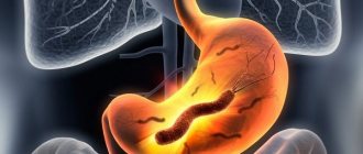
Detection of the bacterium Helicobacter pylori
Previously, the main causes of gastritis were considered to be frequent stress, eating food that irritates the mucous membranes, and irregular nutrition. Only recently have scientists discovered that more than half of the cases are associated with infection. Its danger lies in its oncogenicity - the bacterium can provoke the formation of malignant tumors.
Helicobacter pylori can be detected in the body using the following methods:
- Based on the results of a stool examination. For this purpose, feces are collected in a sterile pharmaceutical container. You cannot take samples from the toilet bowl, as there may be particles of disinfectants there. Feces must be delivered to the laboratory a maximum of 0.5 days after collection. The analysis must be stored in the refrigerator at a temperature no higher than 2 degrees. The results are ready after several hours of waiting and give an accurate answer to the question about the presence of Helicobacter pylori in the body.
- Enzyme immunoassay of venous blood. Detects antibodies to bacteria. If they are detected, a study is carried out using the Western Blot method. In the fluid from the vein, the amount of IgA antibodies to the microbe is determined, thereby establishing the stage of the disease.
Helicobacter pylori lives on the lining of the stomach wall, so it can be found in tissue samples taken during a biopsy. Possibly a breath test. To do this, the patient must drink a liquid with dissolved urea that has a labeled carbon atom. The bacterium quickly breaks down the substance, releasing carbon dioxide. Measuring the level of CO2 in the air that a patient exhales allows the doctor to determine the presence of the microbe.
Conducting pH measurements, x-rays
To make an accurate diagnosis, the doctor must determine the level of stomach acidity. This procedure is called pH measurement. It can be carried out daily or by express method for diagnosing gastritis. For quick analysis, use a probe with an electrode that determines the level of acidity. The daily method tracks indicators throughout the day. This procedure is carried out as follows.
- The probe is inserted through the nose and attached to the patient's belt.
- The patient swallows a special capsule, which is attached to the walls of the stomach.
- Taking tissues during FGDS.
If there are contraindications to the use of a probe, then drugs are used that, under the influence of gastric juice, change the color of urine.
Instrumental methods
For these purposes, various equipment and tools are used. A broader complex is used for chronic inflammation than for acute inflammation. In the latter case, the main method is examination, since the manifestations are more pronounced in children and adults.
FGDS
Fibrogastroduodenoscopy is one of the main diagnostic methods included in the group of endoscopic ones. Tools:
- a probe in the form of a thin flexible tube;
- mini camera on the probe;
- a monitor on which the information received by the camera is visualized.
The examination requires inserting a probe into the digestive organ through the mouth and esophagus. FGDS determines:
- location of inflamed foci in the gastric walls;
- type, nature, stage of membrane damage;
- excludes peptic ulcer disease.
Simultaneously with the stomach, the duodenum is examined, which is often also affected by gastritis. Endoscopic examination results:
- When the mucosa is shiny with fibrin plaque, which is hyperemic and edematous with foci of hemorrhage, non-atrophic or superficial gastritis is diagnosed.
- With severe thinning of the membrane with a smoothed relief, gray color and translucent choroid plexuses, atrophic gastritis is determined. The disease is considered moderate if thinning areas alternate with small areas of white atrophy of varying shapes. If atrophy has reached the last stage, a sharp thinning of the mucous membrane of a cyanotic hue is visualized, which is easy to wound with a simple touch. In this case, folds are not detected.
- With an enlarged pylorus, hyperemic and edematous mucosa, and a significant amount of bile in the stomach, reactive gastritis resulting from chemical poisoning is diagnosed.
- Multiple or single erosive areas form on the mucous membrane during drug-induced gastritis.
- If large folds, similar to the convolutions of the brain, and excessive amounts of mucus are found in the stomach, hypertrophic gastritis is diagnosed. The shell can be easily damaged. Erosions often bleed.
Biopsy
Produced during FGDS. A special probe is used to remove a piece of affected tissue from the walls of the stomach of a child or adult for laboratory testing. The samples are taken from different parts of the stomach. This makes it possible to more accurately determine or refute the presence of Helicobacter, the activity of which is different in different parts of the organ. For this purpose, 2 biopsies are taken from the antrum and the body of the digestive organ. The Helicobacter test is performed using 4 methods:
- bacteriological;
- morphological, which includes the determination of cytology, tissue histology;
- reduced biochemical;
- immunohistochemical.
pH-metry
Standard intragastric pH-metry using the multi-site device “Gastroscan-5”.
It is known that gastritis is an acid-dependent pathology, so an analysis is required to determine the acid content in gastric juice. For these purposes, pH measurement is used, which is classified as:
- Express test is a probe method for measuring acid in the stomach using special electrodes.
- 24-hour pH-metry, which allows you to evaluate the dynamics of fluctuations in acidity levels in two ways:
- tube, carried out by introducing a nasogastric tube into the digestive organ;
- probeless, which involves swallowing a capsule, which is attached to the wall of the stomach to transmit information to the acidogastrometer, and then removed from the body naturally;
- endoscopic pH-metry, which involves taking and analyzing a biopsy sample during FGDS.
Gastric juice assessment
Gastric contents are collected during gastroscopy. Beforehand, an adult or child needs to take a special breakfast with components that stimulate the secretion of digestive juice. The method allows you to determine the cause of inflammation of the walls of the organ. For example, an increased gastrin content indicates the presence of Helicobacter bacteria in the body. Focal gastritis with tissue atrophy is characterized by reduced acidity, decreased activity of pepsin and gastricsin. A strong change in these parameters indicates severe atrophy. Antral gastritis is manifested by three types of secretion:
- hyperreactive;
- hyperparietal;
- panhyperchlorhydric.
But the excess of gastric juice components is not as pronounced as with duodenal ulcerative lesions.
X-ray
Fluoroscopy is performed using a special contrast agent, which the patient must drink before the examination. The method allows you to determine the degree of change in the outline, relief, tone and shape of the organ, detect foci of inflammation and differentiate gastritis from ulcers. It is not recommended for children under three years of age.
Indicative is the double contrast method, which is applicable to children and adults. Barium and air are used as contrast. As the gastrointestinal tract fills, the gastric mucosa carefully straightens, which makes it possible to detect pathology located in the lumen. Additionally, the gastric capacity of the child and adult is assessed.
Detection of Helicobacter pylori
This type of bacterium is the main cause of the development of chronic inflammatory process. The following methods are used for detection:
- checking blood and stool;
- biopsy of affected tissues and cultures taken during FGDS, with further cytological examination of fingerprint smears and histological examination to determine the strain of the bacterium;
- breath test or urea reaction.
The essence of the latter method is to determine the body’s reaction to a solution of urea with a labeled carbon atom that the patient previously drank. If Helicobacter is present in the body, the exhaled air of the patient will contain the product of the breakdown of urea - carbon dioxide - in large quantities.
To confirm the results, a repeat respiratory test is performed after 2 weeks. During the entire course of treatment, the concentration of the microorganism is monitored. After treatment, prevention of gastritis, especially acute gastritis, is required, since there is a high chance of repeated bacterial infection. Therefore, repeat tests are carried out twice a year.
Differentiation
Gastritis must be separated from other pathologies with similar symptoms. For this purpose, a set of methods is used:
- Differentiation of acute forms. Gastritis is separated from acute pancreatitis and cholecystitis by performing a blood test, in which the level of pepsinogens is reduced due to inflammation of the stomach walls. If alpha-amylase levels are elevated, this indicates pancreatitis. With fluoroscopy and FGDS, gastritis is separated from ulcerative lesions of the duodenum or stomach. Based on the ECG result, a gastralgic infarction is determined, which is manifested by the same spasmodic pain in the upper abdomen.
- Differentiation of the chronic form. The symptoms of gastritis are similar to those of ulcers, cancer, neurosis, glandular atrophy of the stomach and secretory dysfunction. To determine cancer, contrast fluoroscopy is performed and a biopsy is taken. With glandular atrophy and secretory dysfunction, changes in the mucosa are not visualized.
X-ray examination
Such an examination will help identify not only gastritis, but also other gastrointestinal pathologies. But the procedure has some contraindications: pregnancy, allergic reactions to active substances and intestinal obstruction. Before diagnosis, the patient must drink liquid barium, iodine or soda solution, which covers the gastric mucosa. How does a doctor define gastritis? The substances stain the stomach, making the walls of the gastrointestinal tract visible on photographs. This examination method is indispensable for urgent hospitalization, since it takes no more than five minutes.
CT scan
This method of diagnosing gastritis is completely safe and is used even by elderly people. Using tomography, you can view the walls of the stomach in detail and identify the presence of formations or deformations on them. Before the procedure, the stomach is filled with inert gas, which allows its walls to straighten. In this way, the elasticity, thickening and narrowing of the walls of the diseased organ or its neoplasms are assessed. The information is recorded in the computer's memory, which allows you to view it again at the right time.
Gastroscopy
This method of examination involves examining the digestive tract in the upper part. Thus, pathologies of the mucous membrane are identified, which are visible on an x-ray. During the procedure, the patient is placed on a couch, then a thin and elastic controlled probe, which is equipped with an optical device - a gastroscope, is inserted into the oral cavity. To avoid the gag reflex, the oral cavity is sprayed with an anesthetic and a special drug is given to drink to reduce discomfort. The procedure does not cause pain. The probe is gradually lowered through the esophagus into the stomach. It stops in the upper part of the small intestine. If during advancement, a specialist can take a biopsy of suspicious tissue with a special instrument that is built into the gastroscope. The sample is sent for histological examination to the laboratory.
The procedure itself takes no more than 30 minutes, but the patient must remain in the hospital after it for about two hours until the medicine stops working. In general, such an examination is not harmful to health, but sometimes bleeding may occur. This can happen due to the formation of perforation in the mucosa, as a result of trauma from the gastroscope. As a rule, after the procedure, a person experiences only painful sensations in the throat, which appear after swallowing the probe.
Probing
This method of diagnosing gastritis is the most popular. The procedure takes about 2.5 hours and is carried out in several stages. Using a special probe, the stomach is tested for its ability to release enzymes into the gastric juice. First of all, the organ is completely emptied using a special tube. Next, 4 portions of basal secretion are collected every 10 minutes. Probing can be performed not only on an empty stomach. If necessary, check the secretion under the influence of food, introduce broth or decoction. Based on the results of the analysis, as well as color, smell and consistency, a diagnosis is made.
So, to detect the disease, you should undergo a full examination by a gastroenterologist, who will conduct an initial examination and tell you what tests to take for gastritis. A detailed examination will help to identify the nature and causes of the disease. Making a correct diagnosis guarantees effective treatment. Therefore, you should not delay going to the doctor.
Preparatory measures
There are factors that can distort test results. To prevent this from happening, you should follow the recommendations:
- A week before the test, it is advisable to stop all medications, especially antibiotics. If this is not possible, then it is imperative to warn the medical staff so that the necessary adjustments can be made to the interpretation of the tests.
- Before collecting stool, you should not eat food containing coarse fiber or coloring pigment (beets, carrots) for three days.
- Fatty, fried, spicy foods should be avoided for 2-3 days; alcohol and smoking.
- Tests are taken in the morning, on an empty stomach. There should be a gap of at least 10 hours between dinner and the collection of biomaterial.
- Immediately before the test, you need to sit quietly and relax for 20-30 minutes. Even slight physical activity can distort the result.
If it is necessary to retake the biomaterial, you must completely repeat the circumstances of the previous test collection: menu, route to the laboratory, etc.
Blood, stool and urine tests are a mandatory part of diagnosing gastritis. Even in the absence of symptoms of the disease, it is necessary to undergo a medical examination once every 6 months if there is a family history of gastrointestinal pathologies, the person leads a sedentary lifestyle, eats incorrectly and irregularly, or abuses smoking or alcohol. Timely treatment and correction of your habits increases the chances of complete relief from gastritis and its complications.
Watch the video on how to quickly identify gastritis:
A blood test for gastritis is an important stage in the diagnosis and treatment of pathology. Therefore, doctors always prescribe a study in order to make an accurate diagnosis. Thanks to this, it is possible to identify the full picture of the disease, as well as find out what stage of the disease the patient currently has.
A blood test is performed on every patient who is admitted to the hospital or for whom precise treatment needs to be prescribed. With the help of a timely study, it is possible to immediately begin therapy for gastritis.